. 2009 Aug 18;42(8):1141-51.
doi: 10.1021/ar900035f.

Soluble polymer carriers for the treatment of cancer: the importance of molecular architecture

Affiliations

Soluble polymer carriers for the treatment of cancer: the importance of molecular architecture

Megan E Fox et al. Acc Chem Res. .

Abstract

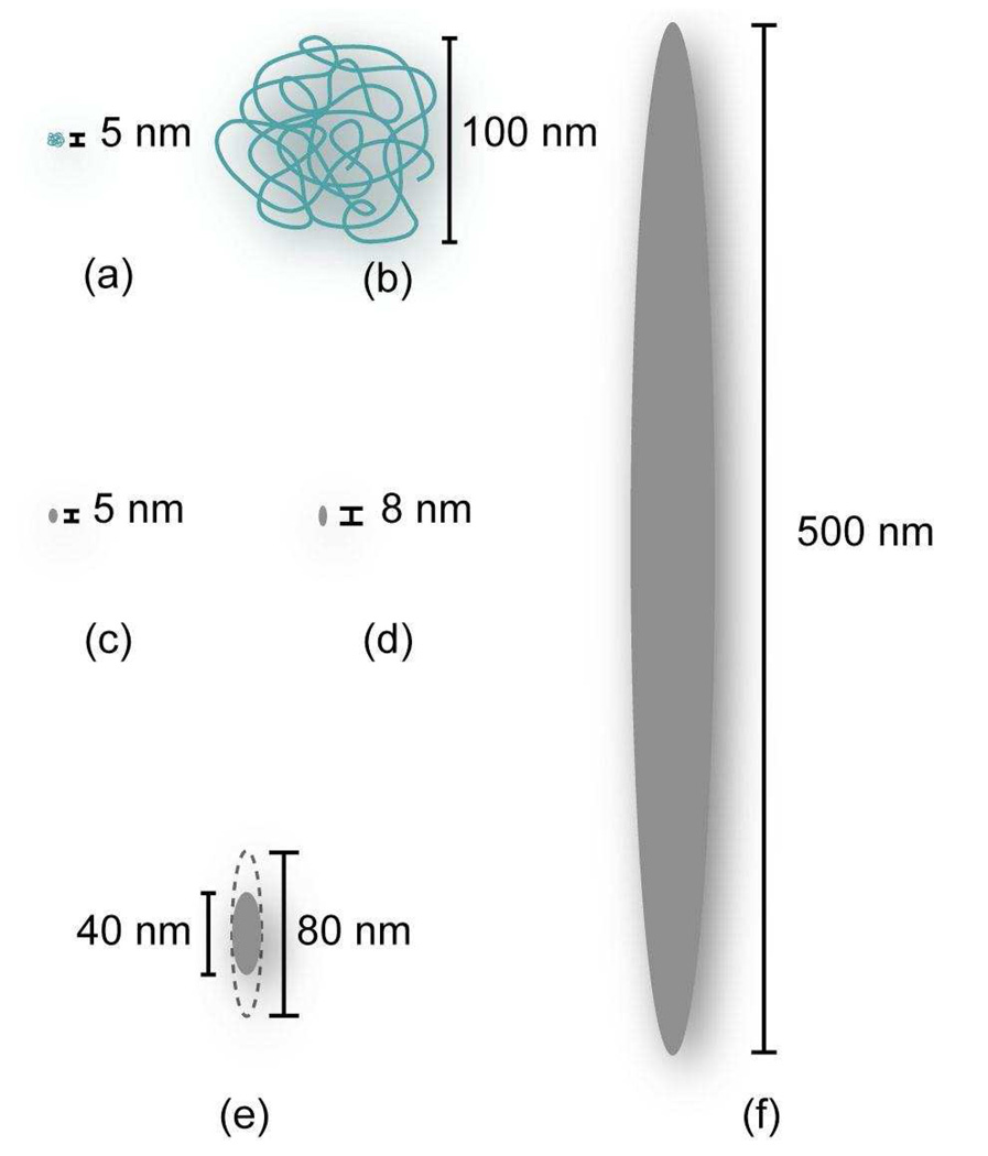
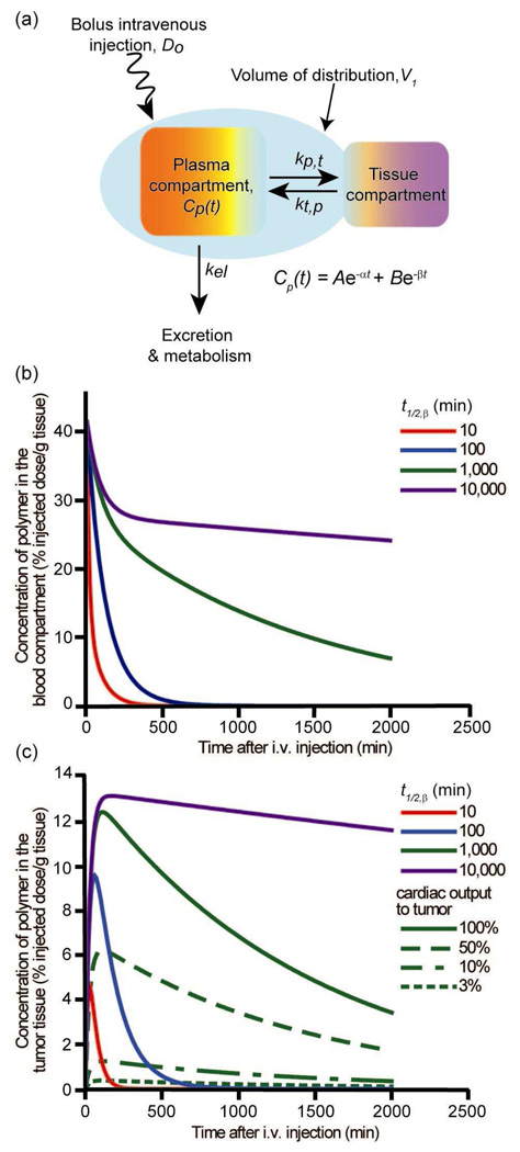
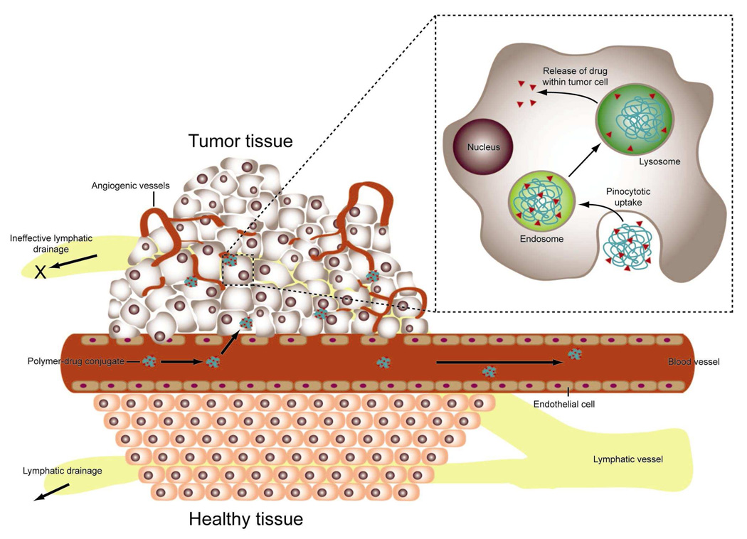
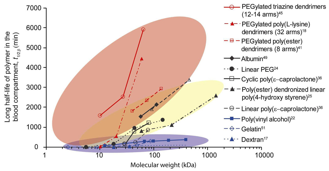
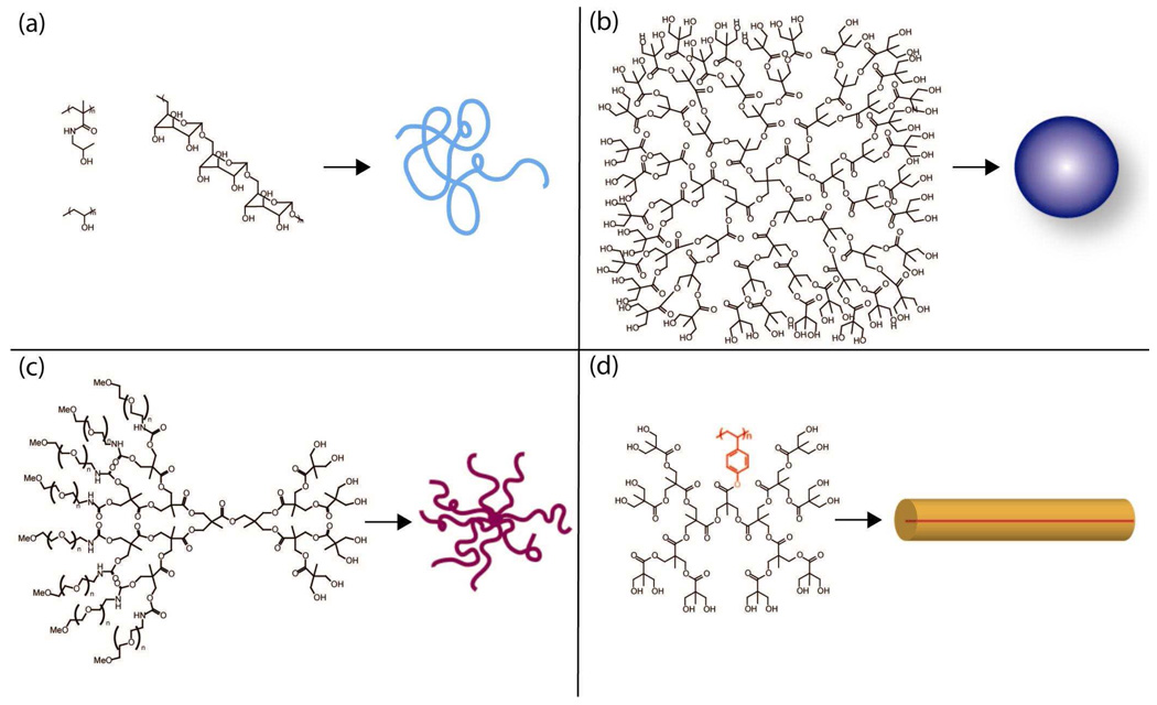
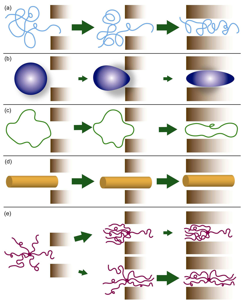
Chemotherapy can destroy tumors and arrest cancer progress. Unfortunately, severe side effects (treatment is usually a series of injections of highly toxic drugs) often restrict the frequency and size of dosages, much to the detriment of tumor inhibition. Most chemotherapeutic drugs have pharmacokinetic profiles with tremendous potential for improvement. Water-soluble polymers offer the potential to increase drug circulation time, improve drug solubility, prolong drug residence time in a tumor, and reduce toxicity. Cytotoxic drugs that are covalently attached to water-soluble polymers via reversible linkages more effectively target tumor tissue than the drugs alone. Macromolecules passively target solid tumor tissue through a combination of reduced renal clearance and exploitation of the enhanced permeation and retention (EPR) effect, which prevails for fast-growing tumors. Effective drug delivery involves a balance between (i) elimination of the polymeric drug conjugate from the bloodstream by the kidneys, liver, and other organs and (ii) movement of the drug out of the blood vasculature and into the tumor (that is, extravasation). Polymers are eliminated in the kidney by filtration through pores with a size comparable to the hydrodynamic diameter of the polymer; in contrast, the openings in the blood vessel structures that traverse tumors are an order of magnitude greater than the diameter of the polymer. Thus, features that may broadly be grouped as the "molecular architecture" of the polymer, such as its hydrodynamic volume (or molecular weight), molecular conformation, chain flexibility, branching, and location of the attached drug, can greatly impact elimination of the polymer from the body through the kidney but have a much smaller effect on the extravasation of the polymer into the tumor. Molecular architecture can in theory be adjusted to assert essentially independent control over elimination and extravasation. Understanding how molecular architecture affects passage of a polymer through a pore is therefore essential for designing polymer drug carriers that are effective in passively delivering a drug payload while conforming to the requirement that the polymers must eventually be eliminated from the body. In this Account, we discuss examples from in vivo studies that demonstrate how polymer architectural features impact the renal filtration of a polymer as well as tumor penetration and tumor accumulation. In brief, features that inhibit passage of a polymer through a pore, such as higher molecular weight, decreased flexibility, and an increased number of polymer chain ends, help prevent elimination of the polymer by the kidneys and can improve blood circulation times and tumor accumulation, thus improving therapeutic effectiveness.


Figures

Figure 1
Figure 1
Relative sizes of two different MW polymers and various pores in the body: (a) PVA chain with MW=13.5kDa (b) PVA chain with MW=580kDa (c) kidney glomerulus pore (d) very large interendothelial junction in healthy tissue (e) typical range of tumor pore diameters (f) very large interendothelial junction in cancerous tissue.
Figure 2
Figure 2
Two-compartment model for predicting polymer concentration in the blood and in the tumor of a mouse (% injected dose/g tissue): (a) schematic representation of the two-compartment model. The concentration of polymer in the blood compartment is given by Cp(t), in which the variables A, B, α, and β are functions of Do, V1, and the rate constants; (b) calculated blood concentration curves versus time. The curves represent polymer concentration profiles for polymers with t1/2,β=10min, 100min, 1,000min, and 10,000min, respectively; (c) solid lines - calculated tumor concentration versus time for polymers with blood t1/2,β=10min, 100min, 1,000min, and 10,000 min, respectively, with 100% of cardiac output passing through the tumor; green lines – calculated tumor concentration versus time for a polymer with t1/2,β=1,000min with different fractions of cardiac output. All calculations assumed mouse with ~2.5g blood, and polymer with kb,t=0.012min−1, kt,b=0.025min−1, and kel=0.0006min−1.
Figure 3
Figure 3
Schematic representation of the EPR effect: passive targeting to tumor tissue is achieved by extravasation of polymers through the increased permeability of the tumor vasculature and ineffective lymphatic drainage. Passively-targeted polymer-drug conjugates are taken up by cancer cells through pinocytosis, and processed by endosomes and lysosomes (Inset). Adapted from ref. .
Figure 4
Figure 4
Effect of MW on blood circulation half-life of intravenously injected polymers for a variety of polymer chemistries and architectures. Red background encompasses polymers with branched and/or globular structures. Yellow background encompasses globular and well-solvated random coil polymers. Blue background encompasses linear polymers that are rapidly cleared from the body. Readers are cautioned to note that the figure is based on reported MW, which does not always scale with actual polymer Vh. Pharmacokinetic experiments for the PEGylated poly(L-lysine) dendrimers and albumin were conducted in normal, healthy rats. The pharmacokinetic data of PVA was collected in tumored mice, however the authors noted that the data was not significantly different from data collected in non-tumored mice. All other data sets were collected from normal, healthy mice.
Figure 5
Figure 5
Structures of polymer drug carriers, and their molecular conformation in solution: (a) linear polymers (HMPA, PVA, dextran) with a loose random coil conformation; (b) Poly(ester) dendrimer with pentaerythritol core (c) PEGylated poly(ester) dendrimers form “star-like” structures with many long arms. (d) Poly(ester) dendronized linear poly(4-hydroxystyrene) forms a tubular “rigid rod” because the backbone is elongated due to the steric requirements of the dendritic branches emanating from the core.
Figure 6
Figure 6
Polymer architecture and the passage of a polymer through a pore for polymers with approximately equivalent Vh: (a) linear random coil polymer readily penetrates and reptates through a pore; (b) polymer with a rigid, globular conformation must deform to pass through; beyond threshold MW both entry and passage through the pore could be difficult; (c) a cyclic polymer lacks a chain end for entering the pore and must deform to enter and pass through it; (d) polymer with a rigid, elongated or tubular conformation easily enters and passes through; (e) arm orientation and distance between chain-ends of branched polymers impact the rate of entry and passage through a pore. While initial entry of only one chain-end (top) may occur rapidly, passage of the entire polymer through the pore is sterically hindered, as the remaining arms must deform for the polymer to pass through; a symmetric conformation (bottom) is less likely since multiple chain ends must penetrate the pore at the same time. Once entry has been achieved passage of the polymer would be less hindered than for the asymmetric distribution.

References

    1. Bader H, Ringsdorf H, Schmidt B. Water-Soluble Polymers in Medicine. Angew. Makromol. Chem. 1984;123/124:457–485.
    1. Kopecek J. Soluble Biomedical Polymers. Polymers in Medicine. 1977;7:191–221. - PubMed
    1. Duncan R. The Dawning Era of Polymer Therapeutics. Nature Reviews Drug Discovery. 2003;2:347–360. - PubMed
    1. Lee CC, Mackay JA, Fréchet JMJ, Szoka FC. Designing Dendrimers for Biological Applications. Nat. Biotechnol. 2005;23:1517–1526. - PubMed
    1. Liu MJ, Fréchet JMJ. Designing Dendrimers for Drug Delivery. Pharmaceutical Science & Technology Today. 1999;2:393–401. - PubMed

Publication types

MeSH terms