. 2009 Oct;35(10):1242-54.
doi: 10.1080/03639040902882280.

Processing difficulties and instability of carbohydrate microneedle arrays

Affiliations

Processing difficulties and instability of carbohydrate microneedle arrays

Ryan F Donnelly et al. Drug Dev Ind Pharm. 2009 Oct.

Abstract

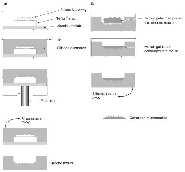
Background: A number of reports have suggested that many of the problems currently associated with the use of microneedle (MN) arrays for transdermal drug delivery could be addressed by using drug-loaded MN arrays prepared by moulding hot melts of carbohydrate materials.

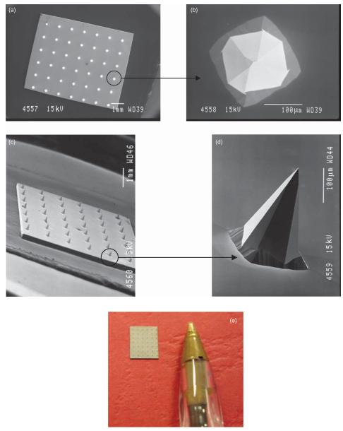
Methods: In this study, we explored the processing, handling, and storage of MN arrays prepared from galactose with a view to clinical application.

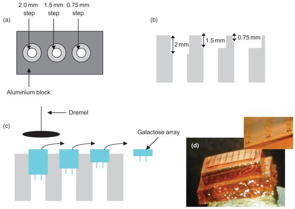
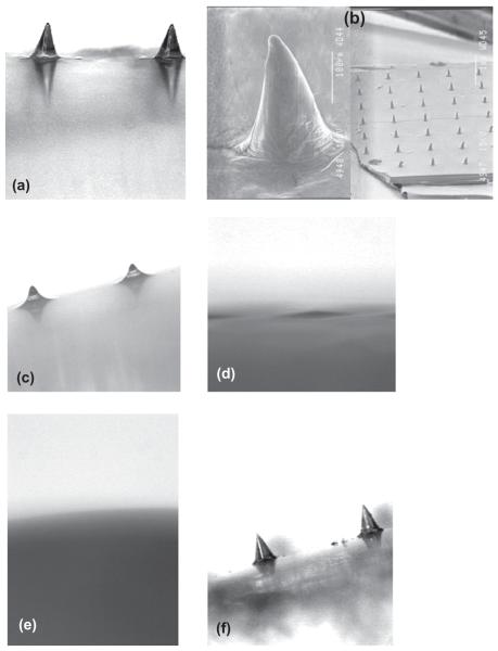
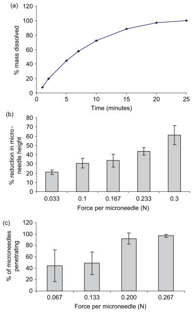
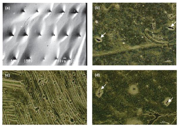
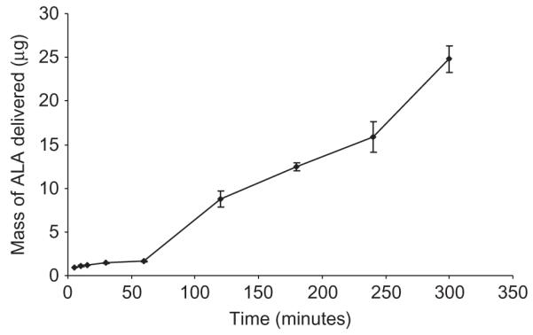
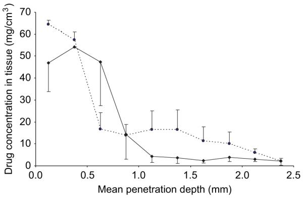
Results: Galactose required a high processing temperature (160 degrees C), and molten galactose was difficult to work with. Substantial losses of the model drugs 5-aminolevulinic acid (ALA) and bovine serum albumin were incurred during processing. While relatively small forces caused significant reductions in MN height when applied to an aluminium block, this was not observed during their relatively facile insertion into heat-stripped epidermis. Drug release experiments using ALA-loaded MN arrays revealed that less than 0.05% of the total drug loading was released across a model silicone membrane. Similarly, only low amounts of ALA (approximately 0.13%) and undetectable amounts of bovine serum albumin were delivered when galactose arrays were combined with aqueous vehicles. Microscopic inspection of the membrane following release studies revealed that no holes could be observed in the membrane, indicating that the partially dissolved galactose sealed the MN-induced holes, thus limiting drug delivery. Indeed, depth penetration studies into excised porcine skin revealed that there was no significant increase in ALA delivery using galactose MN arrays, compared to control (P value < 0.05). Galactose MNs were unstable at ambient relative humidities and became adhesive.

Conclusion: The processing difficulties and instability encountered in this study are likely to preclude successful clinical application of carbohydrate MNs. The findings of this study are of particular importance to those in the pharmaceutical industry involved in the design and formulation of transdermal drug delivery systems based on dissolving MN arrays. It is hoped that we have illustrated conclusively the difficulties inherent in the processing and storage of carbohydrate-based dissolving MNs and that those in the industry will now follow alternative approaches.


Figures

Figure 1
Figure 1
SEM images taken of a typical silicon MN array from directly above (a), of a single MN from above (b), of an array from the side (c), and of an individual MN from the side (d). Digital photograph of a typical silicon MN array (e). For SEM, individual MN arrays were mounted onto aluminium stubs using double-sided adhesive tape and coated in gold (Polaron® E5150 sputter coater; Quorum Technologies, Ringmer, UK). Specimens were then visualized using a JEOL JSM840 scanning electron microscope (Jeol) and images captured on Ilford FP4 black and white roll film (Jessops, Leicester, UK), which was then developed and digitally scanned.
Figure 2
Figure 2
Diagrammatic representation of the steps involved in the preparation of galactose microneedles, showing first production of silicone master moulds from silicon microneedle arrays (a) and second preparation of galactose microneedle arrays from silicone master moulds (b).
Figure 3
Figure 3
Diagrammatic representation of the process of thinning the galactose baseplate. View of the aluminium block from above (a) and from the side (b). Galactose arrays are placed into the aluminium block and ground down using a high-speed abrasive sanding disk fitted to an electric rotary tool (c). Formed galactose microneedle array immediately after completion of micromoulding and prior to baseplate thickness reduction (d).
Figure 4
Figure 4
Light micrograph of galactose microneedles upon preparation (a). Scanning electron micrographs of the same array (b). Light micrograph of galactose microneedles after storage at a relative humidity of 43% for 1 hour (c) and 6 hours (d). Light micrograph of galactose microneedles after storage at a relative humidity of 75% for 1 hour. Light micrograph of galactose microneedles after storage at a relative humidity of 0% for 3 weeks.
Figure 5
Figure 5
The dissolution profile of galactose microneedle arrays in PBS, pH 7.4 (a). Means ± SD, n = 6. Influence of axial loads on height reduction of galactose microneedles (b). Means ± SD, n = 6. Influence of applied forces on the percentage of microneedles penetrating heat-stripped epidermis in vitro (c). Means ± SD, n = 6.
Figure 6
Figure 6
Image showing galactose MN puncturing the Silescol® membrane (a). Holes created in Silescol® membrane upon immediate removal of galactose MNs (b). Silescol® membrane following the 6-hour application of galactose MNs (c). Silescol® membrane 6 hours after the removal of galactose MNs. Image a was taken using a JEOL JSM840 scanning electron microscope (Jeol) and images b, c, and d were obtained using an ECLIPSE TE300 inverted microscope with a DXM1200 digital still camera (Nikon, UK Ltd., Kingston upon Thames Surrey, UK).
Figure 7
Figure 7
Galactose MNs were used to puncture Silescol® membranes, left in place, and 1 mL of a 19 mg/mL solution of ALA was then added to the donor compartment.
Figure 8
Figure 8
ALA penetration into intact (--●--) and MN-punctured —◆—) porcine skin from a 1 mL solution. The formulation was tailored to deliver 19 mg ALA/mL. Galactose MNs were applied for 30 seconds using finger pressure and left in place for 5 hours. Means ± SD, n = 3.

References

    1. Henry S, McAllister DV, Allen MG, Prausnitz MR. Microfabricated microneedles: A novel approach to transdermal drug delivery. J Pharm Sci. 1998;87:922–5. - PubMed
    1. Prausnitz MR. Microneedles for transdermal drug delivery. Adv Drug Deliv Rev. 2004;56:581–87. - PubMed
    1. Cormier M, Johnson B, Ameri M, Nyam K, Libiran L, Zhang DD. Transdermal delivery of desmopressin using a coated microneedle array patch system. J Control Release. 2004;97:503–11. - PubMed
    1. Mikszta JA, Dekker JP, Harvey NG, Dean CH, Brittingham JM, Huang J. Microneedle-based intradermal delivery of the anthrax recombinant protective antigen vaccine. Infect Immun. 2006;74:6806–10. - PMC - PubMed
    1. Coulman SA, Barrow D, Anstey A, Gateley C, Morrissey A, Wilke N. Minimally invasive cutaneous delivery of macromolecules and plasmid DNA via microneedles. Curr Drug Deliv. 2006;3:65–75. - PubMed

Publication types

MeSH terms

LinkOut - more resources