

p63 directly induces expression of Alox12, a regulator of epidermal barrier formation
- PMID: 19555433
- PMCID: PMC2857403
- DOI: 10.1111/j.1600-0625.2009.00894.x
p63 directly induces expression of Alox12, a regulator of epidermal barrier formation
Abstract
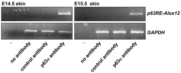
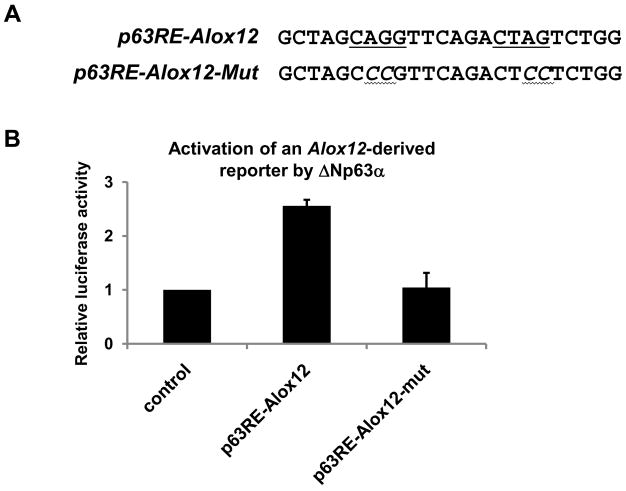
Epidermal development and differentiation are tightly controlled processes that culminate in the formation of the epidermal barrier. A critical regulator of different stages of epidermal development and differentiation is the transcription factor p63. More specifically, we previously demonstrated elsewhere that p63 is required for both the commitment to stratification and the commitment to terminal differentiation. We now demonstrate that DeltaNp63alpha, the predominantly expressed p63 isoform in postnatal epidermis, also plays a role in the final stages of epidermal differentiation, namely the formation of the epidermal barrier. We found that DeltaNp63alpha contributes to epidermal barrier formation by directly inducing expression of ALOX12, a lipoxygenase which contributes to epidermal barrier function. Our data demonstrate that DeltaNp63alpha directly interacts with the promoter of Alox12 in the developing epidermis. Furthermore, we found that the induction of Alox12 expression by DeltaNp63alpha depends on intact p63 binding sites in the Alox12 promoter. Finally, we found that DeltaNp63alpha can induce Alox12 expression only in differentiating keratinocytes, consistent with the role of ALOX12 in epidermal barrier formation.
Figures

Similar articles
-
Dual regulation of TERT activity through transcription and splicing by DeltaNP63alpha.Aging (Albany NY). 2008 Dec 9;1(1):58-67. doi: 10.18632/aging.100003. Aging (Albany NY). 2008. PMID: 20157588 Free PMC article.
-
An active role of the DeltaN isoform of p63 in regulating basal keratin genes K5 and K14 and directing epidermal cell fate.PLoS One. 2009 May 20;4(5):e5623. doi: 10.1371/journal.pone.0005623. PLoS One. 2009. PMID: 19461998 Free PMC article.
-
Differential roles of p63 isoforms in epidermal development: selective genetic complementation in p63 null mice.Cell Death Differ. 2006 Jun;13(6):1037-47. doi: 10.1038/sj.cdd.4401926. Cell Death Differ. 2006. PMID: 16601749
-
Dynamic life of a skin keratinocyte: an intimate tryst with the master regulator p63.Indian J Exp Biol. 2011 Oct;49(10):721-31. Indian J Exp Biol. 2011. PMID: 22013738 Review.
-
P63 deficiency: a failure of lineage commitment or stem cell maintenance?J Investig Dermatol Symp Proc. 2005 Nov;10(2):118-23. doi: 10.1111/j.1087-0024.2005.200416.x. J Investig Dermatol Symp Proc. 2005. PMID: 16363063 Review.
Cited by
-
Interaction of the NRF2 and p63 transcription factors promotes keratinocyte proliferation in the epidermis.Nucleic Acids Res. 2021 Apr 19;49(7):3748-3763. doi: 10.1093/nar/gkab167. Nucleic Acids Res. 2021. PMID: 33764436 Free PMC article.
-
Genetic effects on the skin methylome in healthy older twins.Am J Hum Genet. 2024 Sep 5;111(9):1932-1952. doi: 10.1016/j.ajhg.2024.07.010. Epub 2024 Aug 12. Am J Hum Genet. 2024. PMID: 39137780 Free PMC article.
-
MLL4 mediates differentiation and tumor suppression through ferroptosis.Sci Adv. 2021 Dec 10;7(50):eabj9141. doi: 10.1126/sciadv.abj9141. Epub 2021 Dec 10. Sci Adv. 2021. PMID: 34890228 Free PMC article.
-
Analysis of Skin Regeneration and Barrier-Improvement Efficacy of Polydeoxyribonucleotide Isolated from Panax Ginseng (C.A. Mey.) Adventitious Root.Molecules. 2023 Oct 24;28(21):7240. doi: 10.3390/molecules28217240. Molecules. 2023. PMID: 37959659 Free PMC article.
-
TAp63γ and ΔNp63β promote osteoblastic differentiation of human mesenchymal stem cells: regulation by vitamin D3 Metabolites.PLoS One. 2015 Apr 7;10(4):e0123642. doi: 10.1371/journal.pone.0123642. eCollection 2015. PLoS One. 2015. PMID: 25849854 Free PMC article.
References
-
- Koch PJ, Zhou Z, Roop DR. In: Cornified Envelope and Corneocyte-Lipid Envelope. Elias PM, Feingold KR, editors. Skin Barrier: Marcel Dekker, Inc; 2004. pp. 97–110.
-
- Elias PM, Feingold KR. Lipids and the epidermal water barrier: metabolism, regulation, and pathophysiology. Semin Dermatol. 1992;11:176–182. - PubMed
-
- Segre J. Complex redundancy to build a simple epidermal permeability barrier. Current Opinion in Cell Biology. 2003;15:776–782. - PubMed
-
- Cartlidge P. The epidermal barrier. Seminars in Neonatology. 2000;5:273–280. - PubMed
Publication types
MeSH terms
Substances
Grants and funding
LinkOut - more resources
Full Text Sources
