. 2009 Oct 6;163(2):594-600.
doi: 10.1016/j.neuroscience.2009.06.043. Epub 2009 Jun 23.

Temporal changes in mouse brain fatty acid amide hydrolase activity

Affiliations

Temporal changes in mouse brain fatty acid amide hydrolase activity

S T Glaser et al. Neuroscience. .

Abstract

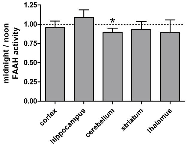
Fatty acid amide hydrolase (FAAH) activity is known to mediate the tone of endogenous fatty acid amides including the endocannabinoid anandamide. FAAH is a potential therapeutic target because genetic or pharmacological ablation of FAAH promotes analgesia and anxiolytic effects without disrupting motor coordination. Little is known about the endogenous temporal fluctuations of brain FAAH activity. This is the first comprehensive study examining temporal fluctuations in mouse brain FAAH activity. Regional mouse brain homogenates were generated at the midpoint of the light ("noon") and dark ("midnight") cycles. While immunoblots revealed no significant changes (P>0.05) in regional activity between these two time points, in vitro activity assays detected a subtle 10% reduction (P<0.05) in cerebellar FAAH activity at midnight. A novel ex vivo autoradiography technique permitted the study of 11 different brain regions, many of which cannot be studied using traditional in vitro methods. The cerebellum and the periaqueductal gray both exhibited significant (P<0.05) reductions in regional FAAH activity in "midnight" brains. These data confirm the need to account for temporal changes in FAAH activity when therapeutically targeting FAAH.


Figures

Fig. 1
Fig. 1. Regional FAAH western blots
Immunoblots for FAAH and actin were run against homogenates generated at the midpoint of the light (“noon”) and dark (“midnight”) cycles.
Fig. 2
Fig. 2. Regional FAAH protein levels
Following sample normalization against actin, FAAH protein levels were compared between noon and midnight conditions. Bars represent the regional mean + standard error. Significance was determined by paired 2-Tailed T-test. No significant (P>0.05) circadian changes were observed in cortical, hippocampal, cerebellar, striatal, or thalamic homogenates.
Fig. 3
Fig. 3. Regional FAAH activity assays
Regional homogenates were generated at the midpoint of the light (“noon”) and dark (“midnight”) cycles, and in vitro FAAH activity assays were performed. Homogenates were incubated at 37°C in buffer containing BSA, 100μM AEA, and [3H]AEA for 15 min. The rate of [3H]ethanolamine production was compared between noon and midnight samples. Significance was determined by paired 2-Tailed T-test. Bars represent the regional mean + standard error. While most regional homogenates generated at noon and midnight exhibited similar rates (P>0.05) of [3H]AEA hydrolysis activity, midnight cerebellar homogenates exhibited a modest, yet significant (★ P<0.05), decline in hydrolysis activity relative to noon samples.
Fig. 4
Fig. 4. Trapping mechanism for ex vivo imaging of FAAH activity
Following the i.v. administration of [3H]AEA, the radiotracer crosses the blood brain barrier. Upon entry into a cell that has active FAAH, [3H]AEA is quickly metabolized into [3H]AA, which is in turn promptly incorporated into membrane phospholipids of the same cell, (as represented by the wide dark grey arrows). This process is mediated by acyl CoA synthetase and acyl transferase and is the predominant metabolic pathway for AA in the rodent brain. As a result, tritium accumulates in cells and brain regions with the most [3H]AEA metabolism. After fifteen minutes, the majority of [3H]AEA hydrolysis and subsequent [3H]AA membrane incorporation is complete, yet the amount of tritium release (as represented by the thin light arrow) from cellular membranes remains negligible , , . For review of both major and alternate arachidonic acid metabolic pathways that must be considered while imaging arachidonic acid, see Rapoport (2003).
Fig. 5
Fig. 5. Regions analyzed by ex vivo autoradiography
A) Serial sections of mouse brains were imaged and the regions depicted in gray were analyzed. They include the 1) caudate putamen, 2) the piriform cortex (cx), 3) the somatosensory (somatosens) cx, 4) the hypothalamus, 5) the amygdala, 6) the thalamus, 7) the hippocampus, 8) the entorhinal cx, 9) the visual cx, 10) the periaqueductal gray (PAG), and 11) the cerebellum. (These images are modified with permission from The Mouse Brain in Stereotaxic Coordinates 28). B) Representative autoradiographs of noon mouse brains.
Fig. 6
Fig. 6. Ex vivo autoradiography assay of FAAH activity
Mice were i.v. administered 1mg/kg AEA + 50μCi [3H]AEA at the midpoint of the light (“noon”) or dark (“midnight”) cycle. Fifteen minutes following tracer administration, brains were isolated, fixed, and processed for imaging in the BetaImager. Regional tritium accumulation (cpm/mm2) due to FAAH activity was quantified using BetaVision software. Regional tritium accumulation was normalized against tritium levels in the pontine nuclei, the brain region with the lowest tritium levels. Normalized regional tritium levels were compared between noon and midnight samples and significance determined by paired 2-Tailed T-test. Bars represent the regional mean + standard error. The dotted line represents the relative value of the pontine nuclei. Midnight cerebellar and PAG exhibited significantly (★ P<0.05) less tritium accumulation relative to noon samples. No other region significantly varied (P>0.05) between noon and midnight samples.

References

    1. Aviello G, Romano B, Izzo AA. Cannabinoids and gastrointestinal motility: animal and human studies. Eur Rev Med Pharmacol Sci. 2008;12(Suppl 1):81–93. - PubMed
    1. Cannon JT, Prieto GJ, Lee A, Liebeskind JC. Evidence for opioid and non-opioid forms of stimulation-produced analgesia in the rat. Brain Res. 1982;243:315–321. - PubMed
    1. Cota D. Role of the endocannabinoid system in energy balance regulation and obesity. Front Horm Res. 2008;36:135–145. - PubMed
    1. Cravatt BF, Demarest K, Patricelli MP, Bracey MH, Giang DK, Martin BR, Lichtman AH. Supersensitivity to anandamide and enhanced endogenous cannabinoid signaling in mice lacking fatty acid amide hydrolase. Proc Natl Acad Sci U S A. 2001;98:9371–9376. - PMC - PubMed
    1. Day TA, Rakhshan F, Deutsch DG, Barker EL. Role of fatty acid amide hydrolase in the transport of the endogenous cannabinoid anandamide. Mol Pharmacol. 2001;59:1369–1375. - PubMed

Publication types