. 2009 Aug 21;284(34):23037-45.
doi: 10.1074/jbc.M109.033332. Epub 2009 Jun 25.

Association of cell surface mucins with galectin-3 contributes to the ocular surface epithelial barrier

Affiliations

Association of cell surface mucins with galectin-3 contributes to the ocular surface epithelial barrier

Pablo Argüeso et al. J Biol Chem. .

Abstract

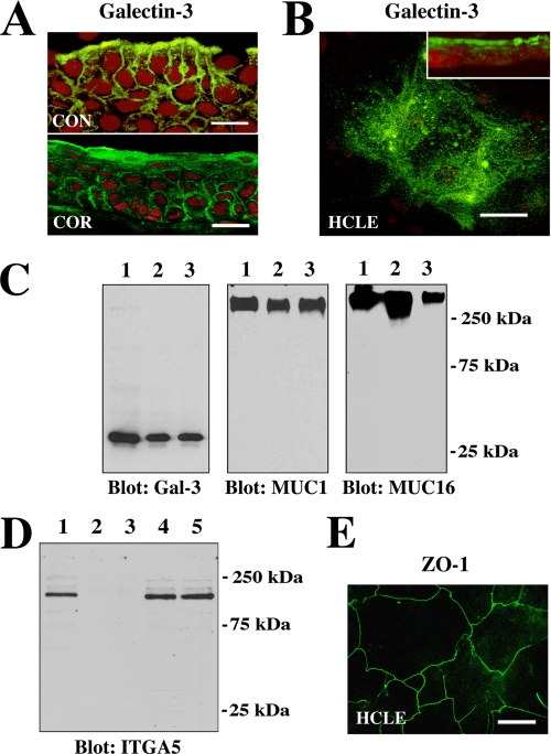
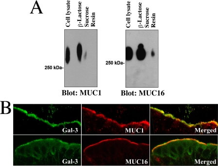
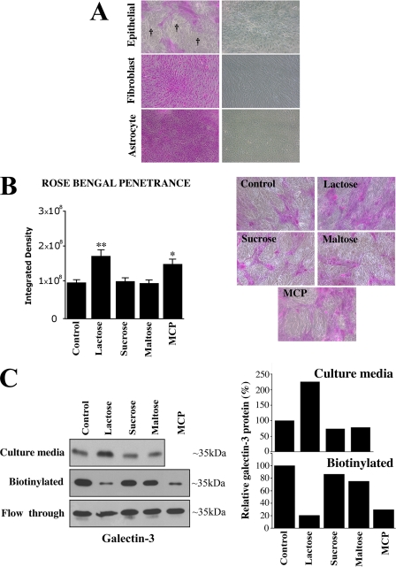
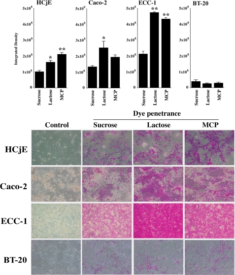
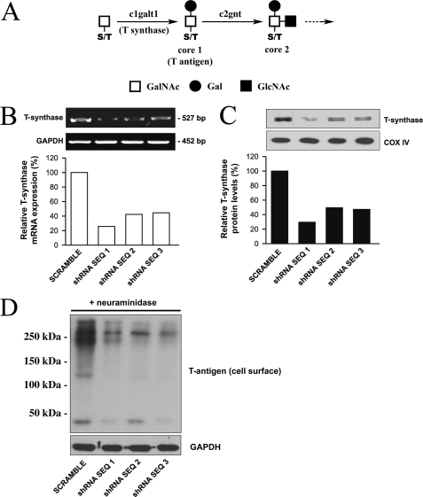
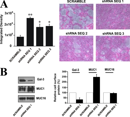
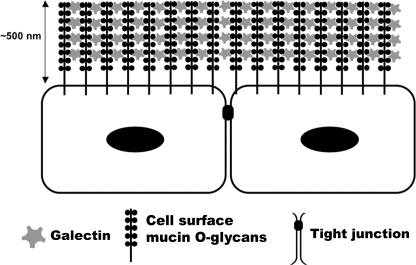
Maintenance of an intact mucosal barrier is critical to preventing damage to and infection of wet-surfaced epithelia. The mechanism of defense has been the subject of much investigation, and there is evidence now implicating O-glycosylated mucins on the epithelial cell surface. Here we investigate a new role for the carbohydrate-binding protein galectin-3 in stabilizing mucosal barriers through its interaction with mucins on the apical glycocalyx. Using the surface of the eye as a model system, we found that galectin-3 colocalized with two distinct membrane-associated mucins, MUC1 and MUC16, on the apical surface of epithelial cells and that both mucins bound to galectin-3 affinity columns in a galactose-dependent manner. Abrogation of the mucin-galectin interaction in four different mucosal epithelial cell types using competitive carbohydrate inhibitors of galectin binding, beta-lactose and modified citrus pectin, resulted in decreased levels of galectin-3 on the cell surface with concomitant loss of barrier function, as indicated by increased permeability to rose bengal diagnostic dye. Similarly, down-regulation of mucin O-glycosylation using a stable tetracycline-inducible RNA interfering system to knockdown c1galt1 (T-synthase), a critical galactosyltransferase required for the synthesis of core 1 O-glycans, resulted in decreased cell surface O-glycosylation, reduced cell surface galectin-3, and increased epithelial permeability. Taken together, these results suggest that galectin-3 plays a key role in maintaining mucosal barrier function through carbohydrate-dependent interactions with cell surface mucins.


Figures

FIGURE 1.
FIGURE 1.
Galectin-3 localizes at the apical surface of normal epithelia in vivo and in vitro. A, confocal immunofluorescence micrograph showing binding of the rat anti-galectin-3 antibody M3/38 (green) to apical membranes of apical epithelial cells in human conjunctival (CON) and corneal (COR) tissue cross-sections. Use of the rabbit anti-galectin-3 antibody H-160 resulted in a similar staining pattern (data not shown). Propidium iodide (red) was used to visualize the cell nuclei. Magnification bars = 20 μm. B, in HCLE cells, binding of the galectin-3 antibody was predominantly apical, as evidenced along the x-y axis (en face view) and in cell culture cross-sections (inset). Magnification bar = 20 μm. C, cell surface proteins on apical cell membranes of HCLE cells were biotinylated, purified through a neutravidin-agarose affinity column, and immunoblotted with anti-galectin-3, anti-MUC1, and anti-MUC16 antibodies. Control lanes were loaded with 15 μg of total protein from HCLE cell lysates (lane 1). Biotinylation experiments were performed in duplicate, and apical proteins were detected in the bound fraction eluted from the affinity column (lanes 2 and 3). D, to determine whether biotinylation was restricted to the apical cell surface, membrane extracts of labeled cells were analyzed for the presence of integrin α5 subunit (ITGA5), a basolateral cell membrane glycoprotein in corneal epithelium (19). In these experiments ITGA5 was not detected in the bound fraction eluted from the neutravidin affinity column (lanes 2 and 3) but was detected in the unfractionated extract (lane 1) as well as in the flow-through fractions (lanes 4 and 5). E, tight junctions in HCLE cells stained with the anti-ZO-1 antibody. The image was acquired along the x-y axis and corresponds to the distribution of tight junctions observed in normal corneal epithelial cells (24). Magnification bar = 50 μm.
FIGURE 2.
FIGURE 2.
Cell surface mucins are counter-receptors for galectin-3 in polarized epithelial cells. A, HCLE cell culture lysates were applied to a galectin-3-agarose affinity column. After extensive washing, the column was eluted with 0.1 m β-lactose. To confirm sugar binding specificity of the galectin-3-binding proteins, the column was eluted with a non-competing disaccharide, 0.1 m sucrose, before elution with β-lactose. The presence of MUC1 (in the corresponding fractions) was detected by Western blot using the 214D4 antibody. β-Lactose effectively eluted MUC1 from the galectin-3 affinity column, indicating that MUC1-galectin-3 interaction was galactose-dependent. In contrast, little MUC1 was detected in the fraction eluted with sucrose. Similar results were obtained when protein blots were probed with OC125 antibody against MUC16, another cell surface-associated mucin expressed in HCLE cells. In these experiments some MUC16 was still detected in the galectin-3 affinity matrix (resin) after elution with β-lactose. B, galectin-3 colocalized with MUC1 and MUC16 in apical membranes of apical cells of the human conjunctival epithelium. Galectin-3 antibody binding was detected with a fluorescein isothiocyanate-conjugated secondary antibody (green), and the mucin antibody binding was detected using a TRITC-conjugated secondary antibody (red). The areas of colocalization of galectin-3 and MUC1 and MUC16 are shown in merged images.
FIGURE 3.
FIGURE 3.
Competitive inhibition of galectin binding disrupts mucosal barrier. A, rose bengal was excluded from islands (†) of differentiated HCLE cells grown for 7 days in the presence of serum, conditions that induce biosynthesis of cell surface mucins and mucin-type O-glycans (21). On the other hand, the dye penetrated into cells that do not express cell surface mucins, such as corneal fibroblasts and brain astrocytes grown under the same conditions. Phase contrast images of cells before dye incubation are shown in the right panel. B, incubation of HCLE cells for 1 h with the galectin binding inhibitors β-lactose and MCP significantly enhanced the penetrance of rose bengal, whereas addition of sucrose, maltose, or media alone (control) did not (*, p < 0.05; **, p < 0.005). Representative micrographs are shown in the right panel. C, galectin-3 was analyzed in culture media and on the cell surface of HCLE cells after incubation for 1 h with galectin binding inhibitors and controls. To determine the presence of galectin-3 in cell culture media, culture supernatants were dialyzed and lyophilized, and non-biotinylated galectin-3 was analyzed by Western blot. To determine the presence of galectin-3 on the cell surface after harvesting the media, cell surface proteins were biotinylated, purified through a neutravidin-agarose affinity column, and immunoblotted with anti-galectin-3. Note the enrichment of non-biotinylated galectin-3 in the cell culture media and the reduction in biotinylated cell surface galectin-3 after incubation β-lactose and MCP. The amount of galectin-3 in the non-biotinylated/neutravidin flow-through did not differ under any of the experimental conditions used in this study. Experiments were performed in duplicate, and the amount of galectin-3 was quantified by densitometry (right) using ImageJ software.
FIGURE 4.
FIGURE 4.
Rose bengal penetrance in normal (HCjE) and neoplastic (Caco-2, ECC-1, BT-20) epithelial cells. Different cell lines of epithelial origin were incubated for 1 h with the non-competitive inhibitor of galectin binding, sucrose, and with the competitive inhibitors, β-lactose and MCP, followed by rose bengal assay. Islands of cells excluding the rose bengal were observed in all epithelial cell lines grown for 7 days after confluence. Competitive inhibition of galactose-dependent surface interactions with β-lactose or MCP significantly increased rose bengal penetrance in HCjE, Caco-2, and ECC-1 cells but not in BT-20, suggesting that the galactose-dependent response of the epithelial cultures is cell type-specific (*, p < 0.01; **, p < 0.001). Representative micrographs are shown in the lower panel.
FIGURE 5.
FIGURE 5.
Abrogation of c1galt1 impairs cell surface O-glycosylation. A, c1galt1 (T-synthase) adds galactose to N-acetylgalactosamine (GalNAc) on serine or threonine (S/T) residues to form core 1 O-glycans in the Golgi complex. Core 1 structures can be further processed by other glycosyltransferases, such as c2gnt that forms the core 2 structure. B, HCLE cells transfected with three shRNA sequences targeting the c1galt1 gene and a scramble control were treated with doxycycline (1 μg/ml). Total RNA (5.0 μg) was analyzed by reverse transcription-PCR. The c1galt1 and glyceraldehyde-3-phosphate dehydrogenase (GAPDH) (loading control) amplifications were performed for 25 and 30 cycles, respectively. Five microliters of PCR mixture were electrophoresed on 1.5% agarose gel and stained by ethidium bromide. Cells transfected with c1galt1 shRNA sequences, had reduced levels of c1galt1 mRNA as compared with cells transfected with scramble shRNA control. C, cell culture lysates collected after treatment with doxycycline (1 μg/ml) were resolved by SDS-PAGE. Ten micrograms of total lysates were subjected to Western blotting using antibodies against T-synthase and cytochrome c oxidase IV (COX IV), the latter serving as loading control. D, to detect the expression level of the T-antigen on cell surface glycoproteins in HCLE cells transfected with c1galt1 shRNA sequences, cell surface proteins were biotinylated and purified through a neutravidin-agarose affinity column, and the eluted material was probed with peanut agglutinin. Before the lectin blot, membranes were pretreated with neuraminidase from Arthrobacter ureafaciens (10 milliunits/ml) in sodium acetate 50 mm, pH 5.0, for 2 h at 37 °C to expose the T-antigen. Cells transfected with c1galt1 shRNA had reduced levels of the T-antigen as compared with scramble control. Glyceraldehyde-3-phosphate dehydrogenase served as a loading control.
FIGURE 6.
FIGURE 6.
C1galt1 contributes substantially to epithelial barrier function. A, treatment of stable HCLE cell lines containing three c1galt1 shRNA sequences with doxycycline (1 μg/ml) for 4 days after reaching confluence resulted in increased permeability to the rose bengal diagnostic dye as compared with the scramble control (*, p < 0.05; **, p < 0.005). Representative micrographs are shown in the right panel. B, C1galt1 knockdown cells contain reduced levels of cell surface galectin-3. Cell surface proteins of HCLE cells transfected with c1galt1 shRNA sequence 1 were biotinylated, purified through a neutravidin-agarose affinity column, and analyzed by Western blot. A decrease in biotinylated galectin-3 was observed in cells transfected with c1galt1 shRNA as compared with the scramble control. In contrast, as expected, there was no reduction in the expression of biotinylated MUC1 and MUC16 cell surface proteins in the c1galt1 knockdown cells. Densitometric values (right) were normalized to scramble control. Experiments were performed in duplicate.
FIGURE 7.
FIGURE 7.
Proposed model of galectin-mucin barrier formation on epithelial surfaces. Our data indicate that two membrane-associated mucins, MUC1 and MUC16, interact with galectin-3 on the epithelial glycocalyx of individual cell membranes to provide a barrier under physiological conditions. Soluble galectin-3 can form multivalent complexes that help to organize mucin assembly on the surface of the cell. The galectin-mucin barrier may regulate the transcellular flux of extracellular components into epithelial cells.

References

    1. Childers N. K., Bruce M. G., McGhee J. R. (1989) Annu. Rev. Microbiol. 43,503–536 - PubMed
    1. Gipson I. K., Argueso P., Beuerman R., Bonini S., Butovich I., Dana R., Dartt D., Gamache D., Ham B., Jumblatt M., Korb D., Kruse F., Ogawa Y., Paulsen F., Stern M., Sweeney D. F., Tiffany J., Ubels J., Willcox M. (2007) Ocul. Surf. 5,179–193 - PubMed
    1. Davies D. E. (2001) Curr. Allergy Asthma Rep. 1,127–133 - PubMed
    1. Einerhand A. W., Renes I. B., Makkink M. K., van der Sluis M., Büller H. A., Dekker J. (2002) Eur. J. Gastroenterol. Hepatol. 14,757–765 - PubMed
    1. Hollingsworth M. A., Swanson B. J. (2004) Nat. Rev. Cancer 4,45–60 - PubMed

Publication types

MeSH terms