Review
. 2009 Jun 26;324(5935):1673-7.
doi: 10.1126/science.1171643.

Growth factors, matrices, and forces combine and control stem cells

Affiliations
Review

Growth factors, matrices, and forces combine and control stem cells

Dennis E Discher et al. Science. .

Abstract

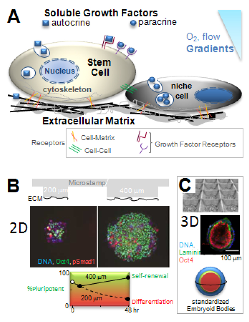
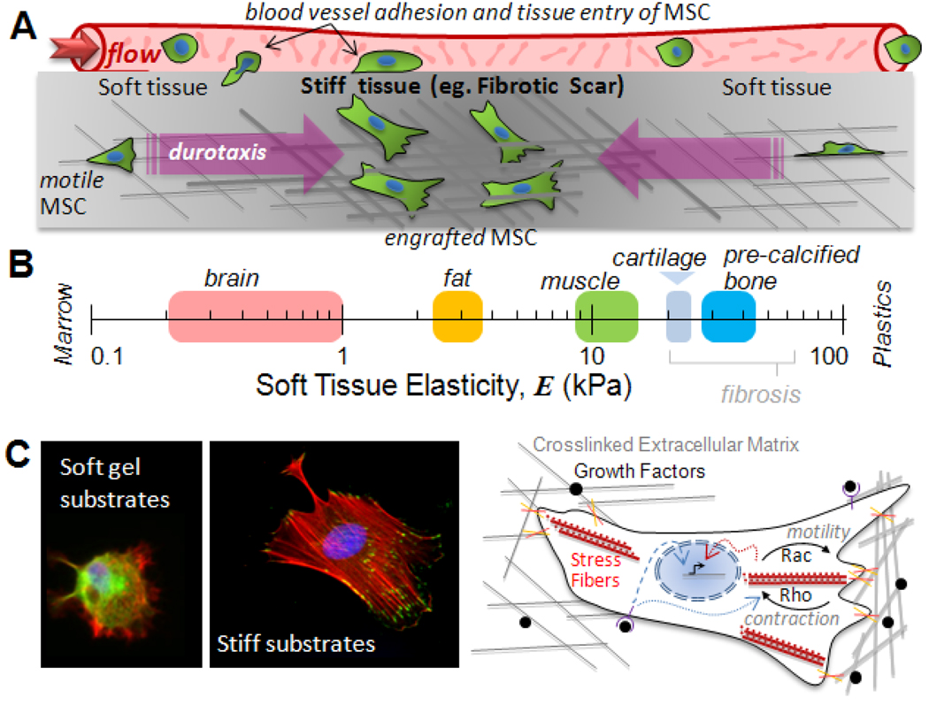
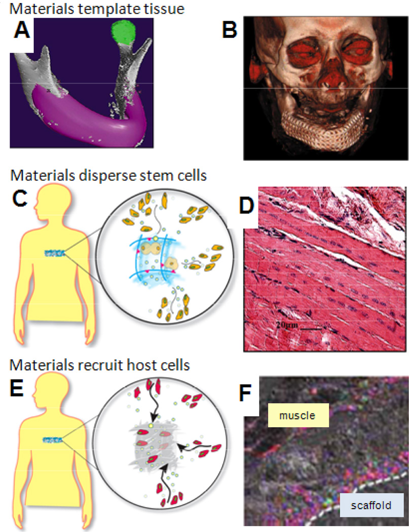
Stem cell fate is influenced by a number of factors and interactions that require robust control for safe and effective regeneration of functional tissue. Coordinated interactions with soluble factors, other cells, and extracellular matrices define a local biochemical and mechanical niche with complex and dynamic regulation that stem cells sense. Decellularized tissue matrices and synthetic polymer niches are being used in the clinic, and they are also beginning to clarify fundamental aspects of how stem cells contribute to homeostasis and repair, for example, at sites of fibrosis. Multifaceted technologies are increasingly required to produce and interrogate cells ex vivo, to build predictive models, and, ultimately, to enhance stem cell integration in vivo for therapeutic benefit.


Figures

Fig 1
Fig 1. Stem cell niche and designs
(A) Soluble and matrix bound factors combine with cell-cell contact, cell-matrix adhesion, and gradients (such as in O2) to direct cell fate. (B) Substrates with 2D micro-patterns of Extracellular Matrix, ECM, control the size of ESC colonies and pluripotency of ESC based on immunofluorescence for the transcription factor Oct4 (15). Plot: large islands increase the pluripotent population that undergoes self-renewal, while small islands inhibit lead to differentiation with a time constant of 24 hr. (C) Substrates with micro-wells help to standardize the diameter of quasi-spherical, 3D embryoid bodies (21). Cells and ECM (laminin) exhibit spherically symmetric pattern, with pluripotent cells in the inside.
Fig 2
Fig 2. Forces in stem cell trafficking
(A) To extravasate from the circulation and invade a tissue, stem cells must adhere strongly to the vessel wall and withstand high flow forces. Within a tissue, additional physical factors can direct motile cells, including durotaxis into stiff, fibrotic regions of tissue where cells engraft. (B) Soft tissue elasticity scale ranging from soft brain (72), fat (73), and striated muscle (25), to stiff cartilage (E ~ 20–30 kPa at the scale of adhesions (74)) and pre-calcified bone (25). (C) In vitro substrates that mimic soft and stiff tissue microenvironments (left) show that cells anchor more strongly to stiff substrates, building focal adhesions and actin-myosin stress fibers. Schematic (right) shows matrix adhesion and growth factors influence both cell physiology and lineage. Signals from growth factor receptors not only propagate into the nucleus (dashed blue arrow) and direct transcription (black arrow), but also affect Rho-GTPase activity (dotted blue arrow). Rac drives motility forward, and Rho regulates contraction of stress fibers (red), and both can also influence gene expression (dotted red arrow).
Fig 3
Fig 3. Synthetic niches in vivo
(A) Materials can fill a specific anatomic defect (pink) to localize transplanted cells and serve as a scaffolding for formation of new tissue. (B) A mandible formed in a patient used a metal and polymer structure that was seeded with MSC and cytokines (49). (C) A designed material niche maintains stem cell viability and proliferation, while promoting outward migration at an appropriate stage of differentiation. (D) Dispersion of stem cells from a niche into regenerating skeletal muscle (60). (E) Recruitment of host stem cells for subsequent homing to sites of tissue injury. (F) Mice with GFP bone marrow-derived cells (green) show regenerating muscle infiltrated with cells that are dual-labeled for endothelial cell marker CD31 (red), indicating neo-vascularization (75).

References

References and Notes

    1. Pittenger M, Martin B. Circ. Res. 2004;95:9. - PubMed
    1. NIH. 2009. http://clinicaltrials.gov.
    1. Alper J. Nat. Biotechnol. 2009;27:213. - PubMed
    1. Macchiarini P, et al. Lancet. 2008;372:2023. - PubMed
    1. Amariglio N, et al. PLoS Med. 2009;6:221.

Additional References in Figure Legends

    1. Flanagan LA, Ju YE, Marg B, Osterfield M, Janmey PA. Neuroreport. 2002;13:2411. - PMC - PubMed
    1. Patel PN, Smith CK, Patrick CW. J Biomed Mater Res A. 2005;73:313. - PubMed
    1. Stolz M, et al. Nat Nanotechnol. 2009;4:186. - PubMed
    1. Fischbach C, et al. Proc Natl Acad Sci U S A. 2009;106:399. - PMC - PubMed

Publication types

Substances