. 2009 Jun 26;324(5935):1726-9.
doi: 10.1126/science.1171716.

Solution nuclear magnetic resonance structure of membrane-integral diacylglycerol kinase

Affiliations

Solution nuclear magnetic resonance structure of membrane-integral diacylglycerol kinase

Wade D Van Horn et al. Science. .

Abstract

Escherichia coli diacylglycerol kinase (DAGK) represents a family of integral membrane enzymes that is unrelated to all other phosphotransferases. We have determined the three-dimensional structure of the DAGK homotrimer with the use of solution nuclear magnetic resonance. The third transmembrane helix from each subunit is domain-swapped with the first and second transmembrane segments from an adjacent subunit. Each of DAGK's three active sites resembles a portico. The cornice of the portico appears to be the determinant of DAGK's lipid substrate specificity and overhangs the site of phosphoryl transfer near the water-membrane interface. Mutations to cysteine that caused severe misfolding were located in or near the active site, indicating a high degree of overlap between sites responsible for folding and for catalysis.


Figures

Figure 1
Figure 1
Structure of diacylglycerol kinase. All panels are for the same representative conformer from the ensemble of structures (fig. S1). Omitted from this figure is the N-terminus (residues 1–25), which was not precisely determined, although it is known to be comprised of two short α-helices that extend over residues 6–14 and 17–23. (A) Ribbon diagram of DAGK with inscribed portico viewed from the membrane plane. DAGK’s five most highly conserved residues are shown (for a single portico only) (B) Ribbon diagram viewed from the membrane plane showing a close-up of the region of the active site containing DAGK’s most highly conserved residues. (C) Ribbon diagram viewed from the cytosol showing side chains for DAGK’s 5 most highly conserved residues (for a single portico only). The connecting loops between the first and second TM segments have been omitted from view so that the organization of the TM helices can more easily be discerned.
Figure 2
Figure 2
Identification of sites critical for catalysis and/or for folding based on the functional analysis of mutants generated by systematically replacing each residue in DAGK with cysteine. (A) Sites labeled blue designate cysteine mutants that exhibited at least 20% of wild type activity. Yellow labels indicate sites for which mutants were observed to fold, but that exhibited less than 20% catalytic activity. Mutation to cysteine at the pink sites resulted in forms of DAGK that were both inactive and misfolded, both in native E. coli membranes and following purification and application of normally-effective refolding protocols. Residues highlighted in the surface-filled representation of DAGK (B) are for a single portico site only (residues are highlighted only for TM1, TM3 and TM2′). Omitted from the surface representations are the imprecisely determined N-termini (residues 1–25). Specific activities for each mutant are listed in table S1. Details of the folding behavior of mutants that exhibit <15% of wild type activity are given in table S2. Sites that are indicated as being conserved with >95% identity were identified based on the multiple sequence alignment presented in figs. S2–S3.
Figure 3
Figure 3
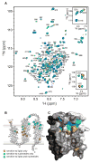
NMR mapping of the substrate binding sites. (A) Example of 800 MHz 1H, 15N-TROSY NMR spectra used to monitor titration of wild type DAGK by a substrate/product or substrate analog. Shown are the overlaid spectra for titration of DAGK by MgAMP-PCP at concentrations of 0 mM (black), 2 mM (red), 4 mM (yellow), 8 mM (green), and 16 mM (blue). Resonances are labeled based on their residue assignments (35). Unlabeled peaks reflect the 10% of resonances for which assignments were not completed. NMR data for titrations of DAGK with DAG, phosphatidic acid, and MgATP are shown in fig. S5. (Insets to A) Selected peaks from the MgAMP-PCP titration (upper panel) and from a separate DAG titration (lower panel, see full spectra in fig. S5). The MgAMP-PCP data illustrate fast exchange NMR conditions in which DAGK peaks gradually change position during the titration, reflecting population-weighted averages between free and complexed forms. In contrast, the data from the DAG titration (lower panel) illustrates slow exchange behavior, where the free enzyme peaks disappear and peaks representing the complex appear at new positions, with contour intensities reflecting the relative populations of the free and complexed states. (B) Summary of active site mapping, highlighting residues associated with TROSY resonances that exhibit large and saturable changes in positions in response to substrate/analog binding. (C) Structure of DAGK with residues highlighted for which TROSY resonances exhibited significant and saturable changes in chemical shift upon binding. In blue are residues that are perturbed by addition of MgATP or MgAMP-PCP (Δδ > 0.02 PPM) and in orange are those perturbed by addition of DAG (Δδ > 0.025 PPM). Residues for which resonance positions are perturbed by both nucleotide and lipid titrations are green.

References

    1. Kennedy EP. Proc Natl Acad Sci U S A. 1982;79:1092. - PMC - PubMed
    1. Raetz CR, Newman KF. J Bacteriol. 1979;137:860. - PMC - PubMed
    1. Badola P, Sanders CR. J Biol Chem. 1997;272:24176. - PubMed
    1. Lis M, Kuramitsu HK. Infect Immun. 2003;71:1938. - PMC - PubMed
    1. Shibata Y, et al. Microbiology. 2009;155:557. - PubMed

Publication types

MeSH terms

Substances

Associated data