. 2009 Oct 1;180(7):657-65.
doi: 10.1164/rccm.200903-0322OC. Epub 2009 Jun 25.

Contribution of epithelial-derived fibroblasts to bleomycin-induced lung fibrosis

Affiliations

Contribution of epithelial-derived fibroblasts to bleomycin-induced lung fibrosis

Harikrishna Tanjore et al. Am J Respir Crit Care Med. .

Abstract

Rationale: Lung fibroblasts are key mediators of fibrosis resulting in accumulation of excessive interstitial collagen and extracellular matrix, but their origins are not well defined.

Objectives: We aimed to elucidate the contribution of lung epithelium-derived fibroblasts via epithelial-mesenchymal transition (EMT) in the intratracheal bleomycin model.

Methods: Primary type II alveolar epithelial cells were cultured from Immortomice and exposed to transforming growth factor-beta(1) and epidermal growth factor. Cell fate reporter mice that permanently mark cells of lung epithelial lineage with beta-galactosidase were developed to study EMT, and bone marrow chimeras expressing green fluorescent protein under the control of the fibroblast-associated S100A4 promoter were generated to examine bone marrow-derived fibroblasts. Mice were given intratracheal bleomycin (0.08 unit). Immunostaining was performed for S100A4, beta-galactosidase, green fluorescent protein, and alpha-smooth muscle actin.

Measurements and main results: In vitro, primary type II alveolar epithelial cells undergo phenotypic changes of EMT when exposed to transforming growth factor-beta(1) and epidermal growth factor with loss of prosurfactant protein C and E-cadherin and gain of S100A4 and type I procollagen. In vivo, using cell fate reporter mice, approximately one-third of S100A4-positive fibroblasts were derived from lung epithelium 2 weeks after bleomycin administration. From bone marrow chimera studies, one-fifth of S100A4-positive fibroblasts were derived from bone marrow at this same time point. Myofibroblasts rarely derived from EMT or bone marrow progenitors.

Conclusions: Both EMT and bone marrow progenitors contribute to S100A4-positive fibroblasts in bleomycin-induced lung fibrosis. However, neither origin is a principal contributor to lung myofibroblasts.


Figures
<b>Figure 1.</b>
Figure 1.
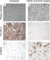
Identification of epithelial–mesenchymal transition (EMT) by primary type II alveolar epithelial cells (AECs) obtained from Immortomice. Cells were cultured in media with or without transforming growth factor (TGF)-β1 (10 ng/ml) and epidermal growth factor (EGF, 100 ng/ml) for 72 hours. With TGF-β1 and EGF treatment, type II AECs (A and B) demonstrated a change to a morphologic appearance consistent with that of fibroblasts on phase-contrast imaging, (C and D) showed reduced expression of the type II AEC protein prosurfactant protein C (pro–SP-C), and (E and F) gained expression of the mesenchymal protein S100A4 as demonstrated by immunocytochemistry.
<b>Figure 2.</b>
Figure 2.
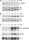
Time course for loss of epithelial markers and gain of mesenchymal markers after treatment with transforming growth factor (TGF)-β1 (10 ng/ml) and epidermal growth factor (EGF, 100 ng/ml). (A and B) Western blots for (A) E-cadherin and (B) S100A4 in Immortomouse type II alveolar epithelial cells (AECs) after treatment with TGF-β1 and EGF. Results were normalized for p44/p42 mitogen-activated protein kinase (MAPK). (C) Northern blot showing increased expression of type I procollagen and S100A4 expression after treatment with TGF-β1 and EGF. Results are normalized for 18S.
<b>Figure 3.</b>
Figure 3.
Cell fate mapping strategy to identify epithelial–mesenchymal transition (EMT) in vivo. (A) Schematic for construction of cell fate reporter mice. (B) 5-Bromo-4-chloro-3-indolyl-β-d-galactopyranoside (X-Gal) staining (blue) in lung sections from untreated R26Rosa.Stop.LacZ.SPC.Cre mice detects reporter gene expression in epithelial cells. (C) X-Gal staining is absent in control (single transgenic) mouse lungs. (D) Epithelial and interstitial cells (arrows) are X-Gal positive 2 weeks after bleomycin administration in lungs of cell fate reporter mice. (BD) Original magnification, ×600.
<b>Figure 4.</b>
Figure 4.
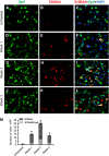
Detection of epithelial–mesenchymal transition (EMT)–derived fibroblasts in lungs after bleomycin treatment. Confocal images to detect fluorescence immunostaining for β-galactosidase (β-Gal) (green), S100A4 (red), and 4′,6-diamidino-2-phenylindole (DAPI) (blue) in lungs from (AC) untreated R26Rosa.Stop.LacZ.SPC.Cre reporter mice and (DL) cell fate reporter mice 1, 2, and 3 weeks after intratracheal bleomycin. Cells that coexpress β-Gal and S100A4 are yellow (arrows point to representative cells). Fluorescence image capture was blanked to sections stained with secondary antibodies only. No green fluorescence was detected in single transgenic mice. (M) Graph representing the number of S100A4+β-Gal+ cells compared with the total S100A4+ cell population in lung parenchyma per high-power field (HPF) from control (untreated) mice and mice 1, 2, and 3 weeks after bleomycin treatment. Mean of 10 HPFs per section ± SEM, n = 3 mice per time point. *P < 0.01 compared with untreated control. (AL) Original magnification, ×600. Control staining images are shown in Figure E2.
<b>Figure 5.</b>
Figure 5.
α-Smooth muscle actin (α-SMA)+ myofibroblasts are rarely derived from epithelial–mesenchymal transition (EMT) after bleomycin treatment. Confocal images to detect fluorescence immunostaining of β-galactosidase (β-Gal)+ cells (green) and α-SMA+ cells (red) and 4′,6-diamidino-2-phenylindole (DAPI) (blue). (AC) Lungs from untreated R26Rosa.Stop.LacZ.SPC.Cre reporter mouse and (DF) reporter mouse 2 weeks after bleomycin treatment. Cells that coexpress β-Gal and α-SMA are yellow (arrow points to representative cell). Dual staining β-Gal+α-SMA+ cells were present but rare in the lung tissue after bleomycin treatment. Weeks 1 and 3 images are not shown but were similar to the Week 2 images. Original magnification, ×600.
<b>Figure 6.</b>
Figure 6.
Few S100A4+ lung fibroblasts coexpress α-smooth muscle actin (α-SMA). Confocal images to detect fluorescence immunostaining for S100A4 (green), α-SMA (red), and 4′,6-diamidino-2-phenylindole (DAPI) (blue) in lungs from (AC) untreated R26Rosa.Stop.LacZ.SPC.Cre reporter mice and (DF) reporter mice 2 weeks after bleomycin treatment. Cells that coexpress S100A4 and α-SMA are yellow (arrow points to representative cell). (G) Bar graph representing the number of S100A4+α-SMA+ cells compared with the total S100A4+ cell population in lung parenchyma per high-power field (HPF) from control (untreated) mice and mice 1, 2, and 3 weeks after bleomycin treatment. Mean of 5 HPF ± SEM, n = 3 mice per time point. *P < 0.01 compared with baseline. (AF) Original magnification, ×600.
<b>Figure 7.</b>
Figure 7.
Bone marrow–derived fibroblasts identified in S100A4.GFP bone marrow chimeras. Top: Immunostaining for (A) S100A4 and (B) green fluorescent protein (GFP; brown staining) in a lung section from a mouse 2 weeks after intratracheal bleomycin administration. (C) Graph showing the number of GFP+ and S100A4+ cells in lung parenchyma per high-power field (HPF) at baseline and 2 weeks after bleomycin treatment. Mean of 10 HPF ± SEM, n = 3 mice per time point. *P < 0.05 compared with untreated baseline. (A and B) Original magnification, ×400.

References

    1. American Thoracic Society, European Respiratory Society. Idiopathic pulmonary fibrosis: diagnosis and treatment [international consensus statement]. Am J Respir Crit Care Med 2000;161:646–664. - PubMed
    1. Phan SH. The myofibroblast in pulmonary fibrosis. Chest 2002;122:286S–289S. - PubMed
    1. Kalluri R, Neilson EG. Epithelial–mesenchymal transition and its implications for fibrosis. J Clin Invest 2003;112:1776–1784. - PMC - PubMed
    1. Zhang K, Rekhter MD, Gordon D, Phan SH. Myofibroblasts and their role in lung collagen gene expression during pulmonary fibrosis: a combined immunohistochemical and in situ hybridization study. Am J Pathol 1994;145:114–125. - PMC - PubMed
    1. Vyalov SL, Gabbiani G, Kapanci Y. Rat alveolar myofibroblasts acquire α-smooth muscle actin expression during bleomycin-induced pulmonary fibrosis. Am J Pathol 1993;143:1754–1765. - PMC - PubMed

Publication types