

Plasma and brain matrix metalloproteinase-9 after acute focal cerebral ischemia in rats
- PMID: 19556529
- PMCID: PMC3712850
- DOI: 10.1161/STROKEAHA.109.554824
Plasma and brain matrix metalloproteinase-9 after acute focal cerebral ischemia in rats
Abstract
Background and purpose: Plasma levels of matrix metalloproteinase-9 (MMP-9) have been proposed to be a useful biomarker for assessing pathological events in brain. Here, we examined the temporal profiles of MMP-9 in blood and brain using a rat model of acute focal cerebral ischemia.
Methods: Plasma and brain levels of MMP-2 and MMP-9 were quantified at 3, 6, 12, and 24 hours after permanent middle cerebral artery occlusion in male Sprague-Dawley rats. Infarct volumes at 24 hours were confirmed with 2,3,5-triphenyl-tetrazolium-chloride staining.
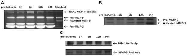
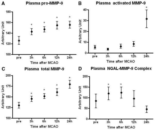
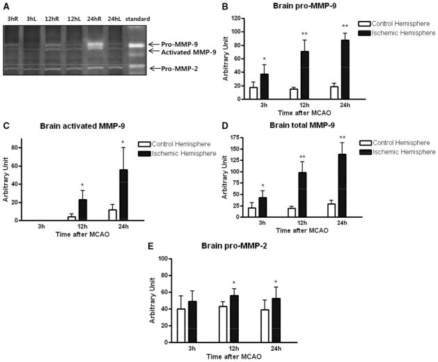
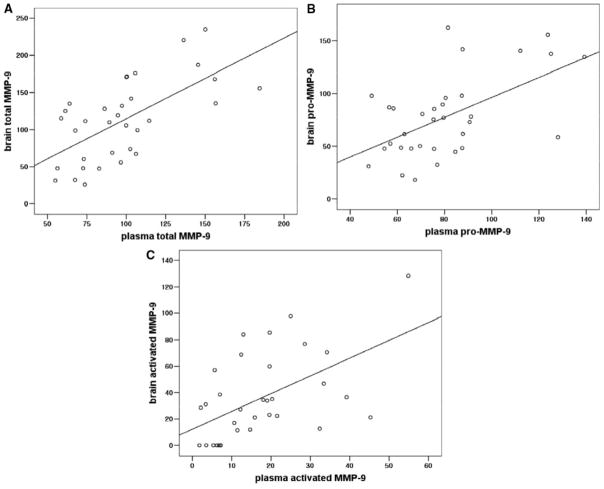
Results: In plasma, zymographic bands were detected between 70 and 95 kDa corresponding to pro-MMP-2, pro-MMP-9, and activated MMP-9. A higher 135-kDa band was also seen that is likely to be NGAL-conjugated MMP-9. After ischemia, there were no significant changes in pro-MMP-2, but plasma levels of pro-MMP-9 steadily increased over the course of 24 hours. Activated MMP-9 levels in plasma were significantly elevated only at 24 hours. Plasma NGAL-MMP-9 complexes showed a transient elevation between 3 to 6 hours, after which levels decreased back down to pre-ischemic baselines. In brain homogenates, pro-MMP-2, pro-MMP-9, and activated MMP-9 were seen but no NGAL-MMP-9 bands were detected. Compared to the contralateral hemisphere, MMP-2 and MMP-9 levels in ischemic brain progressively increased over the course of 24 hours. Overall levels of MMP-9 in plasma and brain were significantly correlated, especially at 24 hours. Plasma levels of pro-MMP-9 at 24 hours were correlated with final infarct volumes.
Conclusions: Elevated plasma levels of MMP-9 appear to be correlated with brain levels within 24 hours of acute cerebral ischemia in rats. Further investigation into clinical profiles of MMP-9 in acute stroke patients may be useful.
Figures
References
-
- Mun-Bryce S, Rosenberg GA. Matrix metalloproteinases in cerebrovascular disease. J Cereb Blood Flow Metab. 1998;18:1163–1172. - PubMed
-
- Yong VW. Metalloproteinases: Mediators of pathology and regeneration in the CNS. Nat Rev Neurosci. 2005;6:931–944. - PubMed
-
- Zhao BQ, Tejima E, Lo EH. Neurovascular proteases in brain injury, hemorrhage and remodeling after stroke. Stroke. 2007;38:748–752. - PubMed
-
- Romanic AM, White RF, Arleth AJ, Ohlstein EH, Barone FC. Matrix metalloproteinase expression increases after cerebral focal ischemia in rats: Inhibition of matrix metalloproteinase-9 reduces infarct size. Stroke. 1998;29:1020–1030. - PubMed
-
- Rosenberg GA, Estrada EY, Dencoff JE. Matrix metalloproteinases and timps are associated with blood-brain barrier opening after reperfusion in rat brain. Stroke. 1998;29:2189–2195. - PubMed
Publication types
MeSH terms
Substances
Grants and funding
LinkOut - more resources
Full Text Sources
Miscellaneous
