. 2009;124(3-4):228-38.
doi: 10.1159/000218128. Epub 2009 Jun 25.

Identification of a maize neocentromere in an oat-maize addition line

Affiliations

Identification of a maize neocentromere in an oat-maize addition line

C N Topp et al. Cytogenet Genome Res. 2009.

Abstract

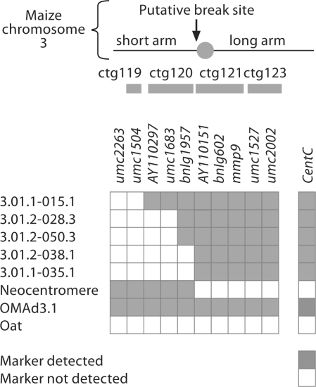
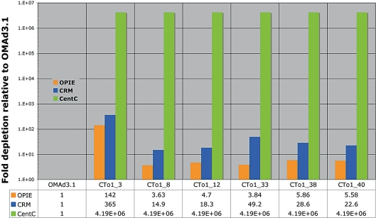
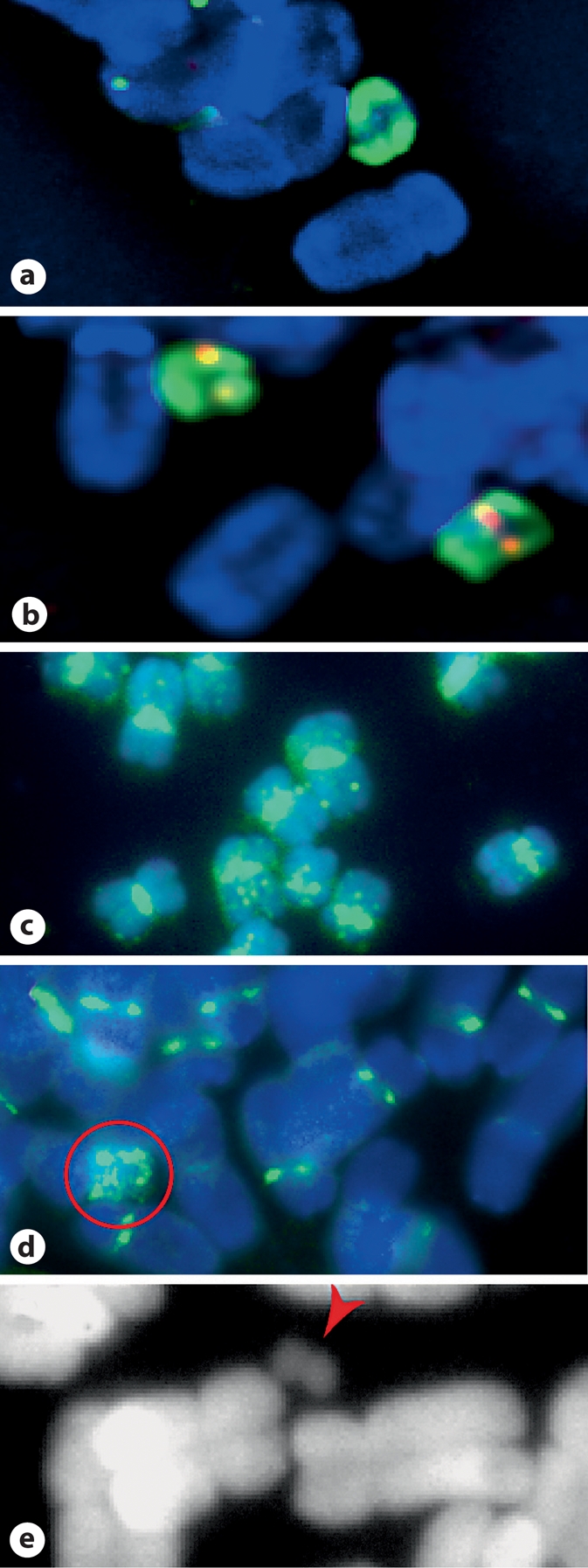
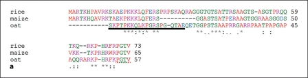
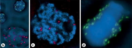
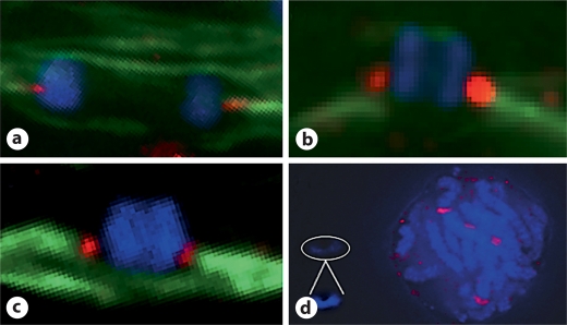
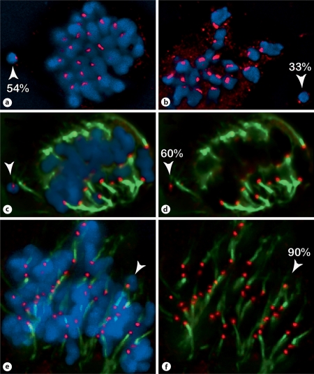
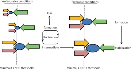
We report a neocentromere event on maize chromosome 3 that occurred due to chromosome breakage. The neocentromere lies on a fragment of the short arm that lacks the primary centromere DNA elements, CentC and CRM. It is transmitted in the genomic background of oat via a new centromere (and kinetochore), as shown by immunolocalization of the oat CENH3 protein. Despite normal transmission of the maize fragment in most progeny, neocentromeres appear to vary in size within the same tissue, as shown by fluorescent measurements. A secondary truncation in one line lowered mitotic transmission to 3% and precipitously reduced the size of the chromosome. The results support the view that neocentromere formation is generally associated with major genomic disturbances such as wide species crosses or deletion of an existing centromere. The data further suggest that new centromeres may undergo a period of instability that is corrected over a period of several generations.


Figures

Fig. 1.
Fig. 1.
Neocentromere material does not contain maize centromere DNA or SSR marker DNA from the maize chromosome 3 long arm. A series of SSR markers spanning maize chromosome 3 were used to probe a panel of chromosome 3 truncation lines, BC1F2–4/9 ‘neocentromere’ progeny, a full maize chromosome 3 disomic addition line (OMAd3.1), and the oat parent of the addition cross (Sun II). The grey bars represent BAC contigs (ctg) that correlate the SSR marker position to the IBM2 physical map. The putative ch3 breakage site maps to the gap between ctg120 and ctg121 on the short arm. Each line was also Southern blotted and probed with the major maize centromere DNA element, CentC, demonstrating the loss of the original centromere in neocentromere lines.
Fig. 2.
Fig. 2.
Maize neocentromere lines lack centromere DNA but are stably inherited. Quantitative PCR analyses of leaf tissue from 6 neocentromere siblings (CTo1_3,8,12,33, 38 and 40) show a sharp decrease in OPIE and CRM retroelements, and did not detect the CentC centromere repeat (Ct = 35 was used arbitrarily for illustration, see Materials and Methods). The full disomic addition line, OMAd3.1 is used as a control. Quantitative measurements presented below the graph are expressed as depletions relative to OMAd3.1 after normalization to the 5S rDNA content of each sample. The maize chromosome of Cto1_3 seems to be inherited at lower frequency than the other neocentromere chromosomes, all of which share the same parent.
Fig. 3.
Fig. 3.
Neocentromere-containing chromosomes are distinguished from oat chromosomes. a DAPI (blue) staining and FISH using an OPIE probe (green) identifies stable neocentromere chromosomes in a metaphase spread, whereas a CentC FISH probe (red) does not stain. b Full addition line chromosomes are larger than their neocentromere counterparts and their native centromeres are identified by CentC staining. c FISH with a CentA probe (green) stains centromeres, pericentromeres, and speckles chromosome arms of maize B73 chromosomes. d The same CentA probe stains oat centromeres/pericentromeres, and stains the maize ch3S neocentromere chromosome asymmetrically (circled in red). e The unstable neocentromere chromosome CTo1_3 (red arrowhead) is a fraction of the stable size (a), and is symmetrical around a primary constriction.
Fig. 4.
Fig. 4.
Identification of the oat CENH3 protein. a A CLUSTAL [Larkin et al., 2007] alignment using the N-termini of the rice, maize, and oat forms of CENH3 proteins. b The underlined sequence was used to generate a peptide antibody that detects oat but not maize CENH3 in mixed cell immunolocalization-FISH experiments. Oat cells are larger and stain for CENH3 (red), whereas maize cells are smaller and stain for CentC (green) but not oat CENH3. c Oat pachytene and d metaphase I immunolocalizations using the oat CENH3 antibody (red or green), stain oat centromeres.
Fig. 4.
Fig. 4.
Identification of the oat CENH3 protein. a A CLUSTAL [Larkin et al., 2007] alignment using the N-termini of the rice, maize, and oat forms of CENH3 proteins. b The underlined sequence was used to generate a peptide antibody that detects oat but not maize CENH3 in mixed cell immunolocalization-FISH experiments. Oat cells are larger and stain for CENH3 (red), whereas maize cells are smaller and stain for CentC (green) but not oat CENH3. c Oat pachytene and d metaphase I immunolocalizations using the oat CENH3 antibody (red or green), stain oat centromeres.
Fig. 5.
Fig. 5.
The maize neocentromere formed submetacentrically. a, b, c CENH3 immunolocalization on meiocytes of stable lines reveal that the neocentromere is located at a submetacentric position on the chromosome arm. CENH3 is shown in red and tubulin is shown in green. d No CENH3 was detected on the unstable CTo1_3 neocentromere from a pachytene cell, despite clear staining on corresponding oat chromosomes (signal intensity of the maize chromosome has been enhanced for viewing – inset).
Fig. 6.
Fig. 6.
CENH3 staining of maize neocentromeres is more variable than staining of established oat centromeres. Cell spreads were immunostained with oat CENH3 (red), tubulin (green, c–f only), and counterstained with DAPI (blue). The fluorescence intensity was measured to estimate kinetochore size. Numbers indicate the relative intensity of the CENH3 of neocentromeres (arrowheads) relative to the mean intensity of oat kinetochores. a, b Two spreads from the same slide illustrating variation in CENH3 intensity. c, d and e, f Two similarly staged cells from the same neocentromere line illustrating a correlation between kinetochore size and attachment to the spindle. The smaller neocentromere (60% of mean oat; c, d) appears to be detached from the spindle, whereas a proportionally much larger neocentromere (90% of mean oat; e, f) is attached to a kinetochore fiber.
Fig. 7.
Fig. 7.
A speculative model of neocentromere establishment. The conditions under which a centromere forms may contribute to its stability. Upon formation, a neocentromere (blue oval) is subject to removal by endogenous forces that normally suppress ectopic centromeres (green arrows). Under certain conditions, reinforcement factors (red and yellow arrows) counteract CENH3 removal and stabilize the neocentromere. If the reinforcement factors are relatively strong (size reflects its relative strength; right panel), establishment is favored and the centromere stabilizes rapidly. If reinforcement factors are weak to begin with (left panel), the centromere may be unstable and fall below the minimal threshold (dotted line). Alternatively, the reinforcement factors may gain strength over time, eventually resulting in a stable centromere.

References

    1. Alonso A, Mahmood R, Li S, Cheung F, Yoda K, Warburton PE. Genomic microarray analysis reveals distinct locations for the CENP-A binding domains in three human chromosome 13q32. Hum Mol Genet. 2003;12:2711–2721. - PubMed
    1. Alonso A, Fritz B, Hasson D, Abrusan G, Cheung F, et al. Co-localization of CENP-C and CENP-H to discontinuous domains of CENP-A chromatin at human neocentromeres. Genome Biol. 8;2007:R148. - PMC - PubMed
    1. Amor DJ, Choo KH. Neocentromeres: Role in human disease, evolution, and centromere study. Am J Hum Genet. 2002;71:695–714. - PMC - PubMed
    1. Amor DJ, Bentley K, Ryan J, Perry J, Wong L, et al. Human centromere repositioning ‘In progress’. Proc Natl Acad Sci USA. 2004;101:6542–6547. - PMC - PubMed
    1. Bhattacharyya GK, Johnson RA. Statistical Concepts and Methods. New York: John Wiley & Sons; 1977. p. 604.

Publication types

MeSH terms