. 2009 Oct;16(10):1362-71.
doi: 10.1038/cdd.2009.79. Epub 2009 Jun 26.

Cell-autonomous requirement for DNaseII in nonapoptotic cell death

Affiliations

Cell-autonomous requirement for DNaseII in nonapoptotic cell death

B P Bass et al. Cell Death Differ. 2009 Oct.

Abstract

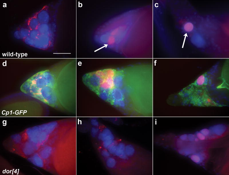
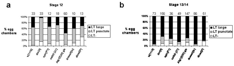
DNA fragmentation is a critical component of apoptosis but it has not been characterized in nonapoptotic forms of cell death, such as necrosis and autophagic cell death. In mammalian apoptosis, caspase-activated DNase cleaves DNA into nucleosomal fragments in dying cells, and subsequently DNase II, an acid nuclease, completes the DNA degradation but acts non-cell autonomously within lysosomes of engulfing cells. Here we examine the requirement for DNases during two examples of programmed cell death (PCD) that occurs in the Drosophila melanogaster ovary, starvation-induced death of mid-stage egg chambers and developmental nurse cell death in late oogenesis. Surprisingly, we found that DNaseII was required cell autonomously in nurse cells during developmental PCD, indicating that it acts within dying cells. Dying nurse cells contain autophagosomes, indicating that autophagy may contribute to these forms of PCD. Furthermore, we provide evidence that developmental nurse cell PCD in late oogenesis shows hallmarks of necrosis. These findings indicate that DNaseII can act cell autonomously to degrade DNA during nonapoptotic cell death.


Figures

Figure 1
Figure 1
DNA fragmentation genes are required for cell death in response to nutrient deprivation. (a) Drawing of an ovariole containing egg chambers at different stages of oogenesis (adapted from 39). FCs are dark gray and NC nuclei in mid-stages are colored black. Nutrient-deprivation induces PCD in early cysts and in mid-oogenesis about st8, whereas developmental NC death occurs between st10 to st14. (b-j) Flies were nutrient-deprived and mid-stage egg chambers were stained with DAPI. (b-e) Progression of cell death in wild-type mid-stage egg chambers. (b) Healthy egg chamber shows dispersed chromatin in NC nuclei. (c) The first signs of degeneration are seen as disorganized clumps of NC chromatin within obvious nuclei. NC nuclei then become highly compact (d) and fragmented (e). (f) At late stages, only small NC remnants are visible (arrow) while FCs are still present and the egg chamber becomes elongated. dICADP homozygous egg chambers show abnormal chromatin compaction (g) and NC nuclei persist until late stages of degeneration (h). (i) DNaseIIlo homozygous egg chambers appear to initiate degeneration normally. (j) In late stages, DNaseIIlo homozygous egg chambers become opaque with dispersed fragments of NC DNA (arrow). All egg chambers are st8-9 and the scale bar is 50μm. All images are projections of 10-16 consecutive images.
Figure 2
Figure 2
DNase II is required for developmental nurse cell programmed cell death. (a-f and k-l) Egg chambers were stained with DAPI and stages were determined by growth of dorsal appendages. (a-c) Progression of developmental NC death in wild-type egg chambers. (a) At st12, the NCs have transferred their cytoplasm to the oocyte and NC nuclei are still large. (b) By st13, some NC nuclei have disappeared and the remaining NC nuclei are condensed. (c) By st14 NC nuclei cannot be detected. (d) A dICADP homozygous st14 egg chamber with a persisting NC nucleus. (e) A DNaseIIlo st14 egg chamber contains NC nuclei and smeared DNA. (f) dICADP; DNaseIIlo double mutant egg chambers resemble DNaseIIlo mutants. (g-h) Egg chambers labeled with TUNEL (green) and DAPI (blue). (g) TUNEL labels some NC nuclei in a wild-type st13 egg chamber (arrows). (h) Reduced TUNEL labeling is seen in dICADP mutant egg chambers (arrow indicates one weakly labeled nucleus). (i) TUNEL labeling is seen around the periphery of NC nuclei in DNaseIIlo homozygous egg chambers (arrow). Long tubular structures in (g) and (h) are dorsal appendages displaying background fluorescence. Egg chambers from DNaseIIlo/Df(3R)sr16 (j) and DNaseIIlo GLC (k) flies show persisting NC nuclei at st14. (l) A degenerating mid-stage egg chamber from a DNaseIIlo GLC shows a less cloudy appearance than that seen in DNaseIIlo homozygotes (Figure 1i). Egg chambers in (a-l) are projections of 10-16 consecutive images. Scale bars are 50μm; images (c-f and j-k) correspond to the scale bar in b and images (g-i and l) correspond to the scale bar in (a).
Figure 3
Figure 3
Lysosome mutants exhibit a disruption in developmental programmed cell death. Persisting NC nuclei are observed in DAPI-stained st14 egg chambers from (a) spinP1 homozygotes, (b) dor4 homozygotes, (c) Atg1Δ3D germline clones, and (d) cathD24 homozygotes. Scale bar is 50μm. Egg chambers are projections of 11-16 consecutive images.
Figure 4
Figure 4
Transient autophagy occurs in dying nurse cells. (a-b) Egg chambers from ovaries of UASp-GFP-Atg8a/NGT; nanos-GAL4/+ flies stained with DAPI. GFP-Atg8a signal can be seen as small puncta in st12 (a), but not st13 (b) egg chambers. Yolk granules within the oocyte also fluoresce. Both images are projections of 12 slices. (c-d) Wild-type st12 and st13 egg chambers (1 μm sections) stained with 1% toluidine blue. N = nucleus; N1-3 are NC nuclei analyzed below. oo = oocyte, DA = dorsal appendage. (e-g) Electron micrographs of organelles in the cytoplasm surrounding NC nucleus 1 (N1) from Figure 4C. FC = Follicle cells, L = lysosome, * = autophagosome. PM = plasma membrane. (h-j) Electron micrographs reveal the progression of nuclear breakdown in NCs. (h) N1 from Figure 4c. Nucleus with compacted chromatin and many surrounding organelles. (i) N2 from Figure 4c. NC with fragmented chromatin and vacuole-like structures. (j) N3 from Figure 4d. Terminal NC with little chromatin remaining. Scale bars in (a-d) are 50 μm. (e-g) at 10,000×, scale bars are 500 nm; (h-j) at 2000×, scale bar is 2 μm.
Figure 5
Figure 5
Lysosome clustering and acidification of nurse cell remnants. All egg chambers stained with DAPI (blue) and LysoTracker (LT, red). (a-c) Progression of LT labeling in wild-type egg chambers. (a) In early st12, increased LT labeling is observed in clusters around NC nuclei. (b) In late st12, acidification of NC nuclei is indicated by large LT-positive domains (arrow). (c) Large LT-positive domains are also observed in st13 egg chambers (arrow). (d-f) Egg chambers from a GFP gene trap in Cp1 (cathepsinL). (d) In early st12, Cp1-GFP clusters around NC nuclei and partially overlaps with LT. (e) In late st12, most Cp1-GFP remains punctate and LysoTracker is detected in large domains. (f) In st13, no significant overlap is detected between CP1-GFP and LT. (g-i) LT labeling in dor4 homozygous mutants reveals a delay in acidification of NC remnants. LT is punctate in st12 (g) and st13 (h) dor4 homozygous egg chambers. (i) A dor4 homozygous st14 egg chamber shows acidification of some persisting NC nuclei. Egg chambers in a-i are projections of 11-21 consecutive images. Scale bar is 50μm.
Figure 6
Figure 6
Quantification of LysoTracker staining. Egg chambers from the genotypes listed on the bottom of each graph were staged and scored for LysoTracker (LT) staining in large domains, punctate spots, or absent/diffuse (LT-). Egg chambers displaying >3 puncta were scored as punctate. Egg chambers displaying both puncta and large domains were scored as having large domains. Separate graphs are shown for st12 (a) and st13/14 (b). Numbers of egg chambers scored are indicated above each column. w1118 was used as a wild-type control.

References

    1. Vaux DL, Korsmeyer SJ. Cell death in development. Cell. 1999;96:245–254. - PubMed
    1. Thompson CB. Apoptosis in the pathogenesis and treatment of disease. Science. 1995;267:1456–1462. - PubMed
    1. Kroemer G, Galluzzi L, Vandenabeele P, Abrams J, Alnemri ES, Baehrecke EH, et al. Classification of cell death: recommendations of the Nomenclature Committee on Cell Death 2009. Cell Death Differ. 2009;16:3–11. - PMC - PubMed
    1. Nagata S. DNA degradation in development and programmed cell death. Annu Rev Immunol. 2005;23:853–875. - PubMed
    1. Parrish JZ, Xue D. Cuts can kill: the roles of apoptotic nucleases in cell death and animal development. Chromosoma. 2006;115:89–97. - PubMed

Publication types

Substances

LinkOut - more resources