. 2009 Jun;5(6):e1000491.
doi: 10.1371/journal.ppat.1000491. Epub 2009 Jun 26.

The structure of a biologically active influenza virus ribonucleoprotein complex

Affiliations

The structure of a biologically active influenza virus ribonucleoprotein complex

Rocío Coloma et al. PLoS Pathog. 2009 Jun.

Abstract

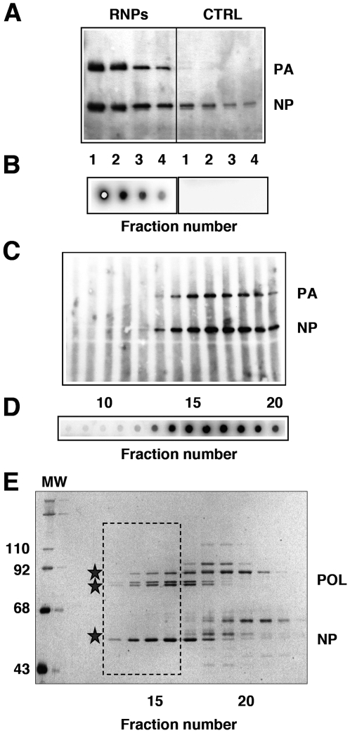
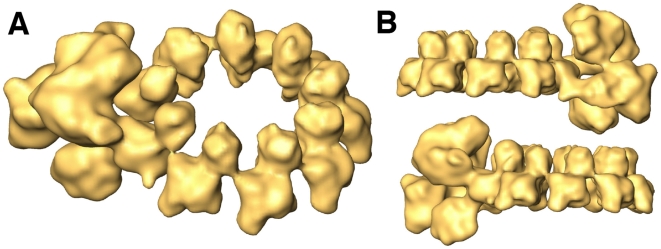
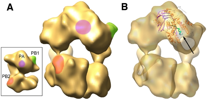
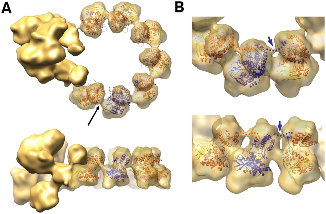
The influenza viruses contain a segmented, single-stranded RNA genome of negative polarity. Each RNA segment is encapsidated by the nucleoprotein and the polymerase complex into ribonucleoprotein particles (RNPs), which are responsible for virus transcription and replication. Despite their importance, information about the structure of these RNPs is scarce. We have determined the three-dimensional structure of a biologically active recombinant RNP by cryo-electron microscopy. The structure shows a nonameric nucleoprotein ring (at 12 Angstrom resolution) with two monomers connected to the polymerase complex (at 18 Angstrom resolution). Docking the atomic structures of the nucleoprotein and polymerase domains, as well as mutational analyses, has allowed us to define the interactions between the functional elements of the RNP and to propose the location of the viral RNA. Our results provide the first model for a functional negative-stranded RNA virus ribonucleoprotein complex. The structure reported here will serve as a framework to generate a quasi-atomic model of the molecular machine responsible for viral RNA synthesis and to test new models for virus RNA replication and transcription.


Conflict of interest statement

The authors have declared that no competing interests exist.

Figures

Figure 1
Figure 1. Generation and purification of a model recombinant RNP.
(A) Recombinant RNPs containing a 248 nt long genomic RNA were generated and amplified in vivo as indicated under Materials and Methods. The RNPs containing a His-tagged PB2 protein (RNPs) were purified by chromatography on Ni-NTA-agarose and the eluted fractions were analysed by Western-blot using anti-PA and anti-NP antibodies. As control, untagged RNPs were generated and purified in parallel (CTRL). (B) The eluted fractions shown in (A) were assayed by in vitro transcription. (C–E) The eluted RNPs were separated on a Sephacryl S300 column and the fractions were analysed again by Western-blot (C) and in vitro transcription as above (D), as well as by silver-staining (E). The position of molecular weight markers (MW) is indicated to the left. The stars indicate the position of the polymerase subunits (POL) and the nucleoprotein (NP). The frame indicates the fractions chosen for electron microscopy analyses.
Figure 2
Figure 2. Three-dimensional model of a recombinant virus mini-RNP.
The final volume is a chimera containing the polymerase and two adjacent NP monomers derived from a non-symmetrical volume and the rest of the NP ring derived from the symmetrical volume (see Fig. S2). (A) Perspective view of the composite three-dimensional structure for the recombinant RNP. (B) Side views.
Figure 3
Figure 3. Docking the atomic structure of PA-PB1 complex into the RNP structure.
(A) The insert shows the three-dimensional model for the virus polymerase complex present in the RNP as reported by Area et al. . The handedness of the structure has been reversed as compared to the one published, as indicated by the docking of the atomic structure of the NP (see Supporting online material). The location of specific domains in the PB1 (green), PB2 (red) and PA (violet) subunits are indicated. A front-view of the polymerase present in the RNP cryo-EM structure is presented, with the locations of the polymerase domains as inferred from the negative-stained model. (B) The same front-view of the polymerase is presented with the docking of the PA(C)-PB1(N) dimer. The N-terminal PB1 peptide is indicated with an arrow and highlighted in green.
Figure 4
Figure 4. Docking the atomic structure of NP monomers into the RNP model.
(A) The atomic structure of NP is represented within the volume of the NP monomers derived from a reconstruction obtained imposing 9-fold symmetry. Upper and side views are presented. One of the monomeric atomic structures is highlighted in blue to reveal the localisation of the connecting loop within the neighbouring monomer. The black arrow points to the potential RNA connection among NP monomers. (B) Close-up view of three NP monomers represented at σ = 1.5. Upper and perspective views are shown. The blue arrow points to the connection between the NP monomers at the top of the molecules. The presumptive connection of the NP head and the loop inserted in the neighbouring monomer is indicated by a dotted line.
Figure 5
Figure 5. Probing the RNP structure by RNAse treatment.
Recombinant RNPs were generated and amplified in vivo as described in Fig. 1. After purification by affinity chromatography on Ni-NTA-agarose, the RNPs were treated with a mixture of pancreatic RNAse (1.2 mg/ml) and T1 RNAse (30 u/ml) for 30 min at room temperature. As a control, the purified RNPs were similarly incubated in the absence of any RNAse. (A) The RNPs were filtered on a Sephacryl S300 column as indicated in Fig. 1 and each fraction was analysed by Western-blot using antibodies specific for NP or PA. The position of NP and PA is indicated to the right. (B) The RNA present in the RNAse-treated or mock-treated RNPs was extracted, terminally labelled with γ-32P-ATP and analysed on a 12% polyacrylamide-urea denaturing gel. Labelled oligonucleotides of 42 and 18 nt in length were run in parallel (M). The mobility of molecular weight markers is indicated to the right.
Figure 6
Figure 6. Phenotype of RNPs with NP mutations in the NP-NP interaction site.
Recombinant RNPs were generated and amplified in vivo using either wt of mutant NP as indicated. After purification of progeny RNPs by pull-down with Ni-NTA-agarose, their accumulation was determined by Western-blot with anti-NP antibodies. (A) Results of a representative experiment, including the analysis of total cell extracts (Input) and the purified RNPs (RNPs). (B) Average and range of two experiments.
Figure 7
Figure 7. Replication in vivo of RNPs containing wt or mutant NP.
The assay for in vivo replication was performed as described in Materials and Methods and the legend to Figure 6. The concentration of RNPs after purification by affinity chromatography was determined by in vitro transcription. (A) Results of a representative experiment, including the analysis of total cell extracts (Input) and the purified RNPs (RNPs). (B) Average and range of two experiments.
Figure 8
Figure 8. Homopolymerisation of wt or mutant NP.
The aggregation state of wt or mutant NPs was determined by gel filtration. Cultures of COS1 cells were transfected with plasmids expressing either wt of mutant NP, as indicated, and total cell extracts were treated with RNAse and filtered on a Sephacryl S300 column. The eluted fractions were analysed by Western-blot with anti-NP antibodies. The position of ferritin (440 kDa) and bovine serum albumin (BSA; 67 kDa) is shown on the top of the Figure.

References

    1. Wright PF, Neumann G, Kawaoka Y. Orthomyxoviridae. In: Knipe DM, Howley PM, editors. Fields Virology 5th edition. Philadelphia: Lippincott Williams & Wilkins; 2007. pp. 1691–1740.
    1. Horimoto T, Kawaoka Y. Influenza: lessons from past pandemics, warnings from current incidents. Nat Rev Microbiol. 2005;3:591–600. - PubMed
    1. Peiris JS, de Jong MD, Guan Y. Avian influenza virus (H5N1): a threat to human health. Clin Microbiol Rev. 2007;20:243–267. - PMC - PubMed
    1. Murti KG, Webster RG, Jones IM. Localization of RNA polymerases of influenza viral ribonucleoproteins by immunogold labeling. Virology. 1988;164:562–566. - PubMed
    1. Palese P, Shaw M. Orthomyxoviridae: the viruses and their replication. In: Knipe DM, Howley PM, editors. Fields Virology 5th edition. Philadelphia: Lippincott Williams & Wilkins; 2006. pp. 1647–1689.

Publication types

MeSH terms