. 2009 Jun;5(6):e1000493.
doi: 10.1371/journal.ppat.1000493. Epub 2009 Jun 26.

Ebola Zaire virus blocks type I interferon production by exploiting the host SUMO modification machinery

Affiliations

Ebola Zaire virus blocks type I interferon production by exploiting the host SUMO modification machinery

Tsung-Hsien Chang et al. PLoS Pathog. 2009 Jun.

Abstract

Ebola Zaire virus is highly pathogenic for humans, with case fatality rates approaching 90% in large outbreaks in Africa. The virus replicates in macrophages and dendritic cells (DCs), suppressing production of type I interferons (IFNs) while inducing the release of large quantities of proinflammatory cytokines. Although the viral VP35 protein has been shown to inhibit IFN responses, the mechanism by which it blocks IFN production has not been fully elucidated. We expressed VP35 from a mouse-adapted variant of Ebola Zaire virus in murine DCs by retroviral gene transfer, and tested for IFN transcription upon Newcastle Disease virus (NDV) infection and toll-like receptor signaling. We found that VP35 inhibited IFN transcription in DCs following these stimuli by disabling the activity of IRF7, a transcription factor required for IFN transcription. By yeast two-hybrid screens and coimmunoprecipitation assays, we found that VP35 interacted with IRF7, Ubc9 and PIAS1. The latter two are the host SUMO E2 enzyme and E3 ligase, respectively. VP35, while not itself a SUMO ligase, increased PIAS1-mediated SUMOylation of IRF7, and repressed Ifn transcription. In contrast, VP35 did not interfere with the activation of NF-kappaB, which is required for induction of many proinflammatory cytokines. Our findings indicate that Ebola Zaire virus exploits the cellular SUMOylation machinery for its advantage and help to explain how the virus overcomes host innate defenses, causing rapidly overwhelming infection to produce a syndrome resembling fulminant septic shock.


Conflict of interest statement

The authors have declared that no competing interests exist.

Figures
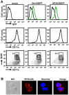
Figure 1
Figure 1. Expression of VP35 in pDCs and cDCs.
A: BMDCs were transduced with pMSCV vector for VP35-EGFP or free EGFP (Ctrl-EGFP) on day 2 and allowed to develop in Flt3L for a total of 8 days. EGFP signals and expression of CD11c and B220 were monitored by flow cytometry. The bracketed areas indicate pDC population and the numbers represent the percentages in the total DC population. B: DCs were transduced with pMSCV vector for VP35-HA. On day 8 the cells were fixed, immunostained with antibody for HA followed by counterstaining with Hoechst for DNA. On the left is an image by differential interference contrast (DIC).
Figure 2
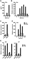
Figure 2. VP35 inhibits induction of IFNα and IFNβ in DCs.
A and B: DCs transduced with VP35-EGFP (VP35, open bar) or free GFP (ctrl, solid bar) were infected with NDV for indicated times. IFNα protein production (A) and Ifnα transcript expression (B) were monitored by ELISA and qRT-PCR, respectively. Values in these experiments (and all below) represent the average of three determinations +/−S.D. C: DCs transduced as above were infected with NDV for 5 h or mock infected and tested for Ifnβ transcripts as in B. D: DCs transduced as above were stimulated with CpG DNA (1 µg/ml) for indicated times and Ifnα transcripts were measured as in B. E: DCs transduced as above or with VP24-EGFP were stimulated with IFNβ (100 U/ml) (solid bar) or vehicle (open bar) for 8 h and expression of Ifit1 and Irf7 transcripts was monitored by qRT-PCR.
Figure 3
Figure 3. VP35 does not inhibit NF-κB activation.
A: DCs transduced with VP35-EGFP (VP35) or free GFP (ctrl) were infected with NDV for indicated times and expression of Tnfα and Ikbα transcripts was monitored as in Figure 2B. B: DCs transduced with VP35-HA or empty vector (ctrl) were infected with NDV for 5 h and immunostained for HA or p65/RelA (NF-κB). About 200 DCs in three different fields were inspected to quantify DCs showing p65 nuclear translocation.
Figure 4
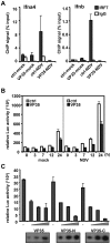
Figure 4. VP35 blocks recruitment of IRF7 to the Ifna and Ifnb genes in DCs.
A: DCs transduced with VP35-HA or with control vector were infected with NDV for 7 h and chromatin was precipitated with anti-IRF7 antibody (solid bar) or normal rabbit IgG (open bar). Precipitated DNA was amplified for the Ifna4 and Ifnb promoters by q-PCR. ChIP signals are expressed as the percentage of input DNA (1%). B: 293T cells (1×10⁵) were transfected with pcDNA3.1 vector for IRF7 (0.05 µg), VP35-HA (VP35) or empty vector (ctrl) (0.2 µg), along with IFNβ luciferase reporter (0.4 µg) and pRL-TK control reporter (0.01 µg) for 24 h, and infected with NDV for 24 h. Reporter activity was monitored by dual luciferase reporter assay. C: A549 cells (1×10⁵) transfected with the vector for Flag-IRF7 (0.02 µg), increasing amounts of VP35-HA (0.2–1 µg) and IFNβ reporter (0.4 µg) plus pRL-TK (0.01 µg) as above were infected with NDV for 24 h and reporter activity was measured as in B. Expression of VP35-HA was verified by immunoblot analysis in the bottom.
Figure 5
Figure 5. Interaction of VP35 with PIAS1 and IRF7.
A: A summary of yeast two-hybrid screen. Two libraries from NDV-stimulated DCs were screened with full length VP35 as a bait. The numbers of sequenced clones are shown. B: 293T cells (1×10⁶) were transfected with pcDNA3.1 vector for full length Flag-PIAS1, or empty vector (2 µg) along with full length VP35-HA (VP35-FL) or truncated versions (2 µg each), for 30 h (see a truncation map on top). Extracts were precipitated by anti-Flag antibody and immunoblotted with anti-HA antibody (top gel). Whole cell extracts (WCE) were blotted with antibody to HA or Flag in the lower gels. C: Cells were transfected with indicated vector for Flag-IRF7 (2 µg) or full length or truncated VP35-HA (2 µg), and extracts were precipitated and blotted as in A. D: Interaction of VP35 with truncated PIAS1 and IRF7. Cells were transfected with HA-tagged full length PIAS1 (FL) (2 µg) or indicated truncations (see a PIAS1 truncation map on top) along with full length VP35-HA and Flag-IRF7 (2 µg each) and extracts were precipitated with anti-Flag antibody, blotted with anti-HA antibody. PIAS1-N migrated just below the Ig heavy chain (marked with *).
Figure 6
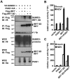
Figure 6. SUMOylation of IRF7 by PIAS1.
A: 293T cells (1×10⁶) were transfected with V5-tagged SUMO3 (0.5 µg), PIAS1-HA (2 µg) along with Flag-IRF7 or Flag-IRF7 6D (1 µg) for 30 h. Extracts were precipitated with antibody to Flag and blotted with antibody to V5. B: Cells were transfected with PIAS1-HA (0.2 µg) or wild type IRF7 alone (0.02 µg) or together, along with IFNβ reporter plus pRL-TK for 24 h followed by NDV infection for 24 h. Luciferase activity was measured as in Figure 4B. C: Above experiments were performed with IRF7 6D in place of wild type IRF7.
Figure 7
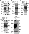
Figure 7. VP35 increases PIAS1 mediated IRF7 SUMOylation.
A: 293T cells (1×10⁶) were transfected with V5-tagged SUMO3 (0.5 µg), Flag-IRF7 (1 µg) along with PIAS1-HA (1 µg) and VP35-HA (2 µg) for 30 h (top panel). Extracts were precipitated with anti-Flag antibody and blotted with anti-V5 antibody. Whole cell extracts were blotted with indicated antibodies (lower panels). B: Cells were transfected with V5-tagged SUMO3 and Flag-IRF7 as above, along with wild type PIAS1-HA or a PIAS1 mutant (PIAS1mu) (1 µg) and extracts were precipitated with anti-Flag antibody and blotted with indicated antibodies. C: Cells were transfected with V5-tagged SUMO3 and Flag-IRF7 as above, along with Ubc9 (1 µg) and extracts were precipitated with anti-Flag antibody and blotted with indicated antibodies. D: 293T cells were transfected with V5-tagged SUMO3 (0.5 µg), Flag-wild type IRF7 or the K406R mutant (1 µg) along with PIAS1-HA (1 µg) and VP35-HA (2 µg) (top panel). Extracts were precipitated with anti-Flag antibody and blotted with anti-V5 antibody. Whole cell extracts were blotted with indicated antibodies (lower panels). E: Cells were transfected with V5-tagged wild type SUMO3 or a conjugation-defective SUMO3 mutant (SUMO3 G/A, see a diagram on the right) (0.5 µg), Flag-tagged wild type IRF7, K406R, or K43R (1 µg) and VP35-HA (2 µg) for 30 h (top panel). Extracts were precipitated with anti-Flag antibody and blotted with anti-V5 antibody. Whole cell extracts were blotted with indicated antibodies (lower panels).
Figure 8
Figure 8. VP35 Increases IRF7 SUMOylation.
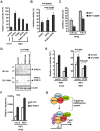
A: Cells were transfected with PIAS1-HA or VP35-HA or both, a vector for IRF7 (0.02 µg each), and IFNβ reporter plus pRL-TK and stimulated with NDV for 24 h, and luciferase activity was measured as in Figure 4B (top panel). B: Experiments above were performed eight times each with triplicate determinations and levels of repression was averaged and quantified. C: Cells were transfected with two doses of VP35 with wild type IRF7 or IRF7 K406R (0.02 µg), along with IFNβ reporter plus pRL-TK for 24 h followed by NDV infection 24 h. Luciferase activity was measured as in Figure 4B. D: L929 cells (1×10⁶) transduced with control or PIAS1 shRNA retroviral vector. Some cells were also transduced with wild type PIAS1-HA or PIAS1r-HA vector that is resistant to PIAS1 shRNA. While cell extracts were blotted with anti-PIAS1 antibody. * denotes a nonspecific band. E: L929 cells (1×10⁵) transduced with control or PIAS1 shRNA vector were transfected with VP35-HA, IRF7 (0.02 µg each) and IFNβ reporter plus pRL-TK and with or without stimulation by NDV for 24 h, and luciferase activity was measured as in Figure 8A. F: NIH3T3 cells (1×10⁵) transduced with control or PIAS1 shRNA vector were further transduced with both of IRF7 and VP35 for 3 days. Cells were then infected with NDV for 24 h and IFNβ transcripts were measured in Figure 2C. G: A model for VP35 action. VP35 interacts with the host SUMOylation machinery, including Ubc9 and PIAS1, the SUMO E2 enzyme and E3 ligase, respectively. VP35 also interacts with IRF7 (and IRF3) bringing IRF7 (and IRF3) to the SUMOylation machinery, and promotes extensive SUMOylation of IRF7 (and IRF3). The premature SUMOylation of the IRFs abrogates their ability to activate IFN transcription causing diminished IFN production. It should be noted (i) VP35 has additional mechanisms of inhibiting IFN transcription and (ii) VP35 may involve other SUMO E3 ligases to increase IRF7 SUMOylation.

References

    1. Feldmann H, Jones S, Klenk HD, Schnittler HJ. Ebola virus: from discovery to vaccine. Nat Rev Immunol. 2003;3:677–685. - PubMed
    1. Bray M, Geisbert TW. Ebola virus: the role of macrophages and dendritic cells in the pathogenesis of Ebola hemorrhagic fever. Int J Biochem Cell Biol. 2005;37:1560–1566. - PubMed
    1. Bosio CM, Aman MJ, Grogan C, Hogan R, Ruthel G, et al. Ebola and Marburg viruses replicate in monocyte-derived dendritic cells without inducing the production of cytokines and full maturation. J Infect Dis. 2003;188:1630–1638. - PubMed
    1. Mahanty S, Hutchinson K, Agarwal S, McRae M, Rollin PE, et al. Cutting edge: impairment of dendritic cells and adaptive immunity by Ebola and Lassa viruses. J Immunol. 2003;170:2797–2801. - PubMed
    1. Geisbert TW, Hensley LE, Larsen T, Young HA, Reed DS, et al. Pathogenesis of Ebola hemorrhagic fever in cynomolgus macaques: evidence that dendritic cells are early and sustained targets of infection. Am J Pathol. 2003;163:2347–2370. - PMC - PubMed

Publication types

MeSH terms