Review
. 2009;53(5-6):795-804.
doi: 10.1387/ijdb.072483jr.

Pattern formation in the Drosophila eye disc

Affiliations
Review

Pattern formation in the Drosophila eye disc

Jean-Yves Roignant et al. Int J Dev Biol. 2009.

Abstract

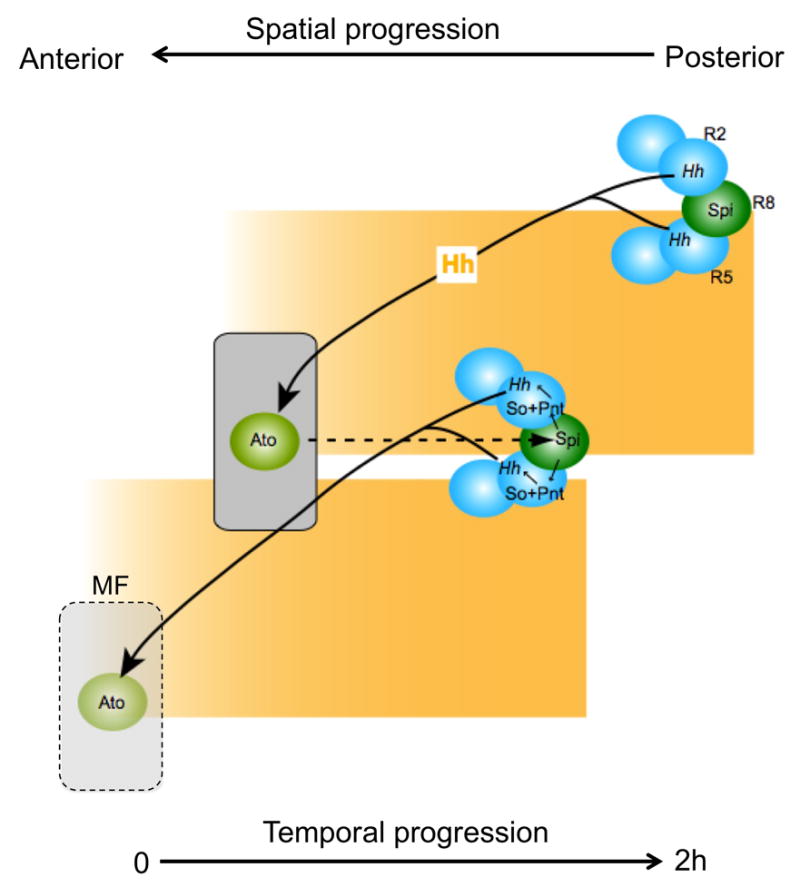
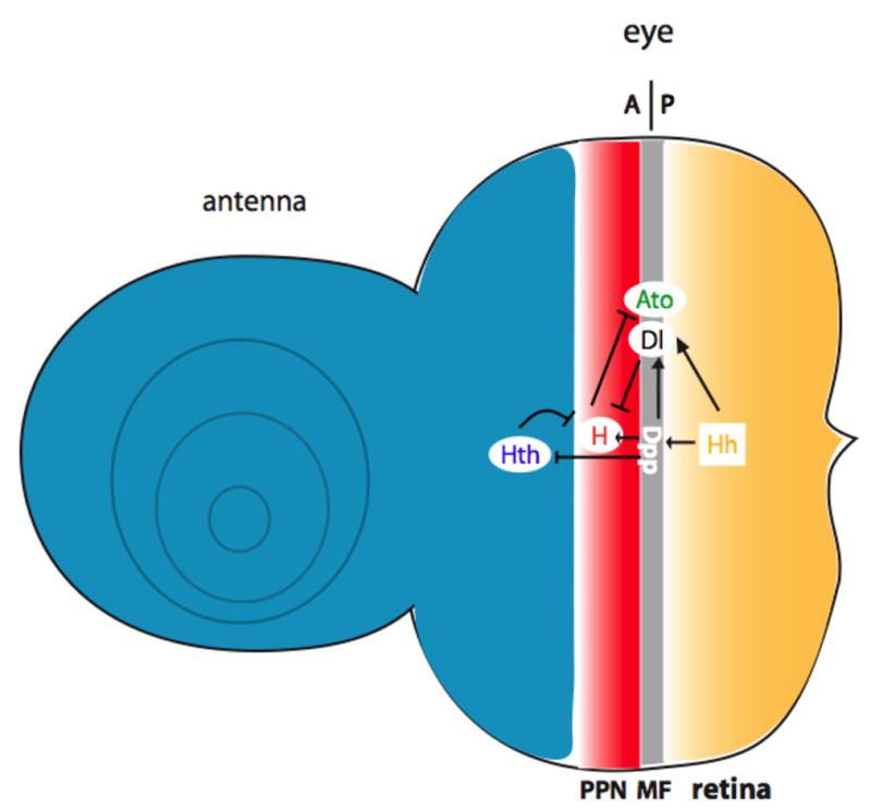
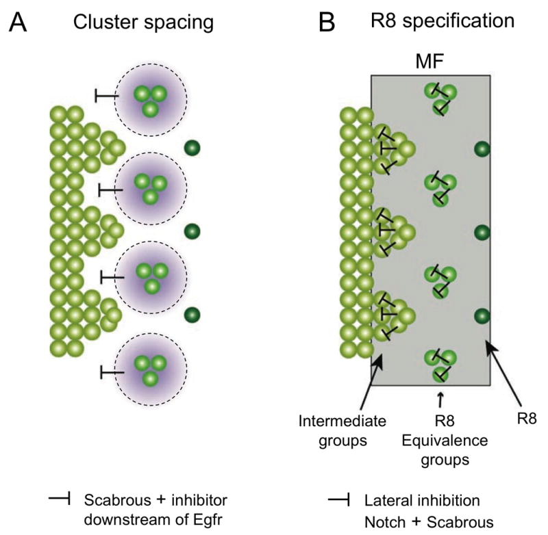
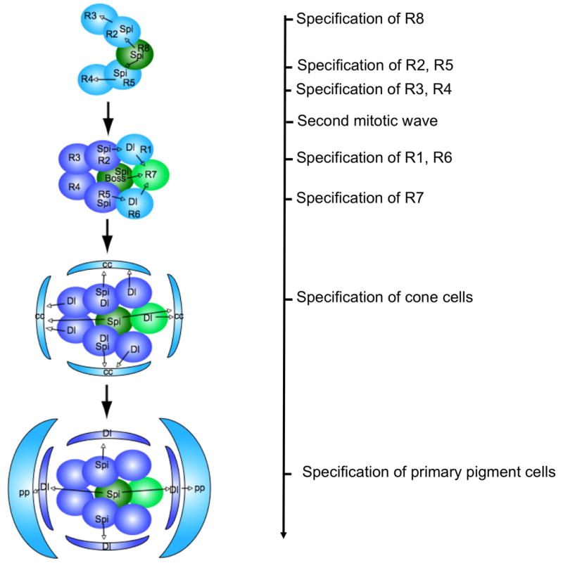
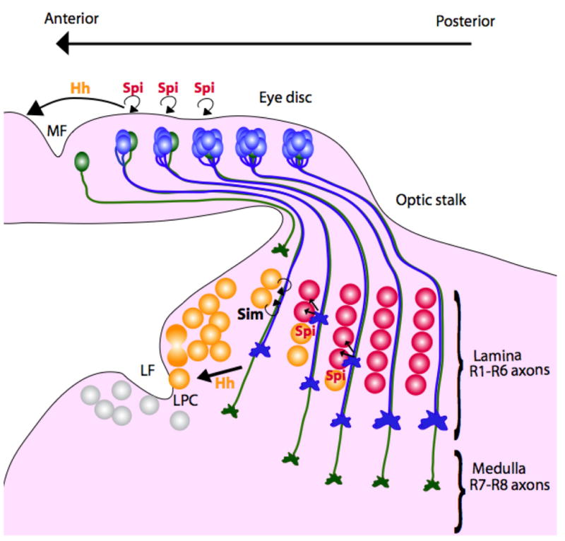
Differentiation of the Drosophila compound eye from the eye imaginal disc is a progressive process: columns of cells successively differentiate in a posterior to anterior sequence, clusters of cells form at regularly spaced intervals within each column, and individual photoreceptors differentiate in a defined order within each cluster. The progression of differentiation across the eye disc is driven by a positive autoregulatory loop of expression of the secreted molecule Hedgehog, which is temporally delayed by the intercalation of a second signal, Spitz. Hedgehog refines the spatial position at which each column initiates its differentiation by inducing secondary signals that act over different ranges to control the expression of positive and negative regulators. The position of clusters within each column is controlled by secreted inhibitory signals from clusters in the preceding column, and a single founder neuron, R8, is singled out within each cluster by Notch-mediated lateral inhibition. R8 then sequentially recruits surrounding cells to differentiate by producing a short-range signal, Spitz, which induces a secondary short-range signal, Delta. Intrinsic transcription factors act in combination with these two signals to produce cell-type diversity within the ommatidium. The Hedgehog and Spitz signals are transported along the photoreceptor axons and reused within the brain as long-range and local cues to trigger the differentiation and assembly of target neurons.


Figures

Figure 1
Figure 1. A delayed autoregulatory loop of Hh expression drives MF progression
Hh is secreted by differentiating photoreceptors, primarily R2 and R5, and diffuses anteriorly to activate ato expression in the MF. Ato then promotes R8 specification and expression of the proteases Rho and Ru, which cleave the Spi precursor to produce active Spi. Spi acts locally to enhance the activity of the transcription factor Pnt, which together with So activates an eye-specific enhancer of the hh gene. The resulting Hh expression reiterates the cycle, which repeats approximately every 2 h in a spatial progression from posterior to anterior.
Figure 2
Figure 2. Hh induces long-range and short-range secondary signals that control the precise position of the MF
Hh acts over a short range to induce the expression of Dpp, which diffuses over a long range to turn off hth and turn on hairy, establishing a preproneural domain (PPN). Hh and Dpp also induce the expression of Delta, a transmembrane ligand that acts on adjacent cells to turn off hairy and allow ato expression, initiating photoreceptor differentiation.
Figure 3
Figure 3. Inhibitory signals control cluster spacing and R8 specification
(A) shows that spacing of intermediate groups is controlled by inhibitory signals from clusters in the preceding column, including Sca and a second factor downstream of EGFR signaling. (B) shows the process of restriction of Ato expression within the MF from a continuous stripe of cells to intermediate groups, 3-cell R8 equivalence groups, and single R8 cells. R8 selection requires local lateral inhibition mediated by Notch and Sca.
Figure 4
Figure 4. Sequential recruitment of ommatidial cells is controlled by the short-range ligands Spi and Dl
Spi promotes the differentiation of photoreceptors R2, 5, 3, 4, 1 and 6. It also induces the expression of Dl, which acts together with Spi and Boss to promote R7 differentiation, together with Spi to promote cone cell differentiation, and alone to promote primary pigment cell differentiation. Cells are added sequentially because Spi and Dl can only act over a short distance.
Figure 5
Figure 5. Hh and Spi signals are transported down the photoreceptor axons to organize the target region
Hh promotes the final division of lamina precursor cells (LPC) posterior to the lamina furrow (LF), and through Sim, their association with photoreceptor axons. Hh also induces the LPCs to express Dac, which activates expression of the EGFR, allowing them to respond to Spi. Cells that respond to Hh by expressing Dac and EGFR are colored yellow. Spi then promotes LPC differentiation adjacent to retinal axons. Cells that respond to Spi by expressing Elav are colored red. Release of the same signals from the cell body and the axons coordinates morphogenesis of the eye and the lamina.

Comment in

  • Pattern formation today.
    Chuong CM, Richardson MK. Chuong CM, et al. Int J Dev Biol. 2009;53(5-6):653-8. doi: 10.1387/ijdb.082594cc. Int J Dev Biol. 2009. PMID: 19557673 Free PMC article. Review.

Similar articles

Cited by

References

    1. APIDIANAKIS Y, GRBAVEC D, STIFANI S, DELIDAKIS C. Groucho mediates a ci-independent mechanism of hedgehog repression in the anterior wing pouch. Development. 2001;128:4361–70. - PubMed
    1. ARTAVANIS-TSAKONAS S, RAND MD, LAKE RJ. Notch signaling: Cell fate control and signal integration in development. Science. 1999;284:770–6. - PubMed
    1. AZA-BLANC P, RAMIREZ-WEBER FA, LAGET MP, SCHWARTZ C, KORNBERG TB. Proteolysis that is inhibited by Hedgehog targets Cubitus interruptus protein to the nucleus and converts it to a repressor. Cell. 1997;89:1043–53. - PubMed
    1. BAKER NE, MLODZIK M, RUBIN GM. Spacing differentiation in the developing Drosophila eye: A fibrinogen-related lateral inhibitor encoded by scabrous. Science. 1990;250:1370–7. - PubMed
    1. BAKER NE, YU S, HAN D. Evolution of proneural atonal expression during distinct regulatory phases in the developing Drosophila eye. Curr Biol. 1996;6:1290–301. - PubMed

Publication types

MeSH terms

LinkOut - more resources