

Reptile scale paradigm: Evo-Devo, pattern formation and regeneration
- PMID: 19557687
- PMCID: PMC2874329
- DOI: 10.1387/ijdb.072556cc
Reptile scale paradigm: Evo-Devo, pattern formation and regeneration
Abstract
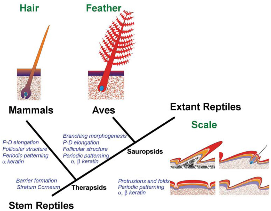
The purpose of this perspective is to highlight the merit of the reptile integument as an experimental model. Reptiles represent the first amniotes. From stem reptiles, extant reptiles, birds and mammals have evolved. Mammal hairs and feathers evolved from Therapsid and Sauropsid reptiles, respectively. The early reptilian integument had to adapt to the challenges of terrestrial life, developing a multi-layered stratum corneum capable of barrier function and ultraviolet protection. For better mechanical protection, diverse reptilian scale types have evolved. The evolution of endothermy has driven the convergent evolution of hair and feather follicles: both form multiple localized growth units with stem cells and transient amplifying cells protected in the proximal follicle. This topological arrangement allows them to elongate, molt and regenerate without structural constraints. Another unique feature of reptile skin is the exquisite arrangement of scales and pigment patterns, making them testable models for mechanisms of pattern formation. Since they face the constant threat of damage on land, different strategies were developed to accommodate skin homeostasis and regeneration. Temporally, they can be under continuous renewal or sloughing cycles. Spatially, they can be diffuse or form discrete localized growth units (follicles). To understand how gene regulatory networks evolved to produce increasingly complex ectodermal organs, we have to study how prototypic scale-forming pathways in reptiles are modulated to produce appendage novelties. Despite the fact that there are numerous studies of reptile scales, molecular analyses have lagged behind. Here, we underscore how further development of this novel experimental model will be valuable in filling the gaps of our understanding of the Evo-Devo of amniote integuments.
Figures
Comment in
-
Pattern formation today.Int J Dev Biol. 2009;53(5-6):653-8. doi: 10.1387/ijdb.082594cc. Int J Dev Biol. 2009. PMID: 19557673 Free PMC article. Review.
References
-
- Alexander NJ. Comparison of α an β keratin in reptiles. Z. Zellforsch. 1970;110:153–165. - PubMed
-
- Alibardi L. Keratohyalin-like granules and differentiation of the shedding complex in embryonic and regenerating epidermis of lizards and Sphenodon (Reptilia, Lepidosauria) Amphibia-Reptilia. 1999;20:11–23.
-
- Alibardi L. Ultrastructural localization of alpha-keratins in the regenerating epidermis of the lizard Podarcis muralis during formation of the shedding layer. Tissue Cell. 2000;32:153–162. - PubMed
Publication types
MeSH terms
Grants and funding
LinkOut - more resources
Full Text Sources
Other Literature Sources
