. 2009 Sep;28(9):435-42.
doi: 10.1089/dna.2008.0850.

Uptake of foreign nucleic acids in kidney tubular epithelial cells deficient in proapoptotic endonucleases

Affiliations

Uptake of foreign nucleic acids in kidney tubular epithelial cells deficient in proapoptotic endonucleases

Timea Buzder et al. DNA Cell Biol. 2009 Sep.

Abstract

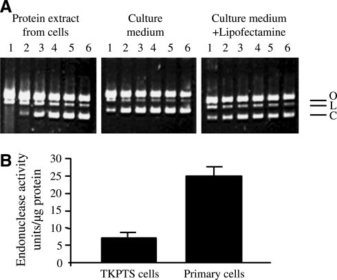
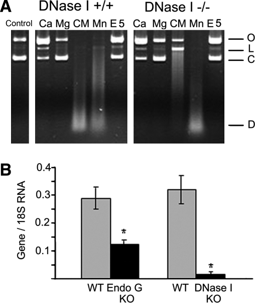
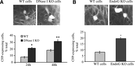
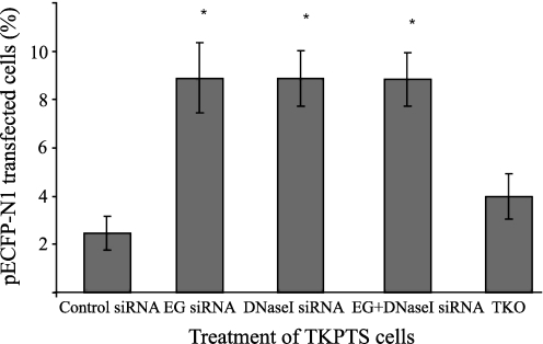
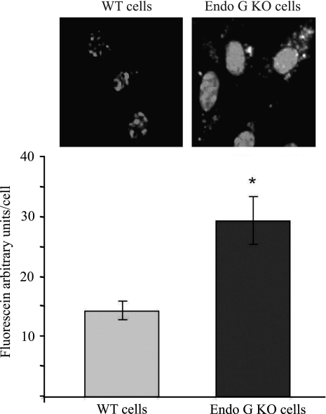
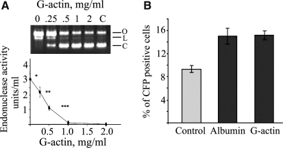
Degradation of DNA during gene delivery is an obstacle for gene transfer and for gene therapy. DNases play a major role in degrading foreign DNA. However, which of the DNases are involved and whether their inactivation can improve gene delivery have not been studied. We have recently identified deoxyribonuclease I (DNase I) and endonuclease G (EndoG) as the major degradative enzymes in the mouse kidney proximal tubule epithelial (TKPTS) cells. In this study, we used immortalized mouse TKPTS cells and primary tubular epithelial cells isolated from DNase I or EndoG knockout (KO) mice and examined the degradation of plasmid DNA during its uptake. DNase I and EndoG KO cells showed a higher rate of transfection by pECFP-N1 plasmid than wild-type cells. In addition, EndoG KO cells prevented the uptake of fluorescent-labeled RNA. Complete inhibition of secreted DNase I by G-actin did not improve plasmid transfection, indicating that only intracellular DNase I affects DNA stability. Data demonstrate the importance of DNase I and EndoG in host cell defense against gene and RNA delivery to renal tubular epithelial cells in vitro.


Figures

FIG. 1.
FIG. 1.
Endonuclease activity in tubular epithelial cell extract and culture medium. The activity was measured using pBR322 PIA as described in the Materials and Methods section. (A) Endonuclease activity is present both in the cellular protein extracts and in the culture medium (left and middle panels). Pretreatment with Lipofectamine does not protect plasmid DNA against in vitro digestion by endonucleases (right panel). Dilutions (1–6) of cell extract or medium 1:1, 1:5, 1:25, 1:125, 1:625, and 1:3125, respectively. O, open circular DNA (with one or more single-strand DNA breaks but no double-strand breaks); L, linear DNA (with one double-strand DNA break); C, covalently closed circular DNA (without DNA breaks), which is the primary substrate for endonucleases. Endonuclease activity is seen only in the first two dilutions in cell extract, and in nondiluted culture medium. (B) Endonuclease activity in immortalized TKPTS cells and PTE cells. Primary cells have a higher total endonuclease activity (25 ± 2 units/μg protein in primary cells vs. 7 ± 3 units/μg protein in TKPTS cells, n = 3–6, p < 0.001) as measured using the pBR322 PIA in the presence of Ca2+ and Mg2+ ions (2 mM CaCl2 and 5 mM MgCl2), which are the cofactors for most of the cellular endonucleases. PIA, plasmid incision assay; TKPTS, mouse kidney proximal tubule epithelial; PTE, primary tubular epithelial.
FIG. 2.
FIG. 2.
Activity and expression of endonucleases in PTE cells. (A) In the total protein extracts isolated from DNase I WT mice, the strongest endonuclease activity could be obtained when Ca2+ and Mg2+ ions were added together, resulting in digested DNA. This is characteristic to DNase I, which therefore provides most of the endonuclease activity in the normal kidney. In the kidney tissue extracts obtained from DNase I KO mice, Mn-dependent endonuclease was the most prominent, suggesting that EndoG is the second major endonuclease in the absence of DNase I (Widlak et al., 2001). Vertical row: O, open circular DNA; L, linear DNA; C, covalently closed circular DNA; D, digested DNA. Horizontal row: control nondigested pBR322 DNA; Ca2+, 2 mM CaCl2, pH 7.5; Mg2+, 2 mM MgCl2, pH 7.5; CM [Ca2++Mg2+], 2 mM CaCl2 + 2 mM MgCl2; Mn2+, 2 mM MnCl2, pH 7.5; E5, 2 mM EDTA, no cations, pH 5 (to measure DNase II activity). (B) Expression of endonucleases in WT, EndoG KO, and DNase I KO cells measured using real-time reverse transcriptase polymerase chain reaction. DNase I expression is wiped out almost completely, while EndoG KO is partially inhibited because these cells were isolated from heterozygous animals (n = 4, *p < 0.001). DNase I, deoxyribonuclease I; WT, wild-type; KO, knockout; EndoG, endonuclease G.
FIG. 3.
FIG. 3.
Efficiency of plasmid transfection of PTE cells with active or inactive endonucleases. (A) Expression of CFP after pECFP-N1 plasmid transfection in DNase I KO PTE cells is higher than in WT cells. KO versus WT (21 ± 5% vs. 8 ± 5% transfected cells, n = 3, *p < 0.013; 32 ± 6% vs. 18 ± 5% transfected cells, n = 3, **p < 0.025). Two time-points have been used, 24 h and 48 h incubation after transfection. The second time-point shows higher transfection efficiency and has been chosen for experiment B, where EndoG WT and KO cells have been transfected with the same plasmid. (B) Expression of CFP 48 h after pECFP plasmid transfection in EndoG KO PTE cells is higher than in WT cells (19 ± 5% vs. 8 ± 5% CFP-positive cells, n = 6, *p < 0.001). CFP, cyan fluorescent protein.
FIG. 4.
FIG. 4.
Efficiency of pECFP-N1 plasmid transfection in TKPTS cells after EG siRNA, DNase I siRNA, and EG+DNase I siRNA treatment. TKPTS cells were treated with EndoG, DNase I, and EndoG+DNase I siRNA using TransIT-TKO transfection reagent (TKO), controlled with siCONTROL Non-Targeting siRNA (control siRNA) and transfected with pECFP-N1 cyan plasmid. SiRNA-silenced cells (EG, DNase I and both) show significantly higher plasmid transfection efficiency (8.8% vs. 2.4% of pECFP-N1 transfected cells, *p = 0.001). siRNA, small interfering RNA.
FIG. 5.
FIG. 5.
Efficiency of RNA transfection of PTE cells with active or inactive EndoG. Primary EndoG KO or WT mouse tubular epithelial cells were transfected with fluorescent siRNA as described in the Materials and Methods section. About 48 h later RNA transfection was detected using a fluorescent microscope. Blue color of DAPI was used to stain the nuclei. EndoG KO cells show significantly higher rate of siRNA transfection than WT cells (14 ± 2 vs. 29 ± 4 arbitrary fluorescence units per cell, n = 3, *p < 0.01).
FIG. 6.
FIG. 6.
Extracellular DNase I does not influence DNA transfection efficiency. (A) TKPTS cells were treated with different concentration of G-actin in culture medium, and endonuclease activity was measured with PIA. G-actin inhibited DNase I in culture medium (n = 4 per concentration point, *p = 0.018, **p = 0.0052, ***p = 0.006). (B) The efficiency of TKPTS cells transfection with pECFP plasmid in the presence of 1 mg/mL G-actin was not different from the one measured in the presence of 1 mg/mL albumin (15 ± 5 vs. 15 ± 8% of CFP-positive cells, n = 6).

References

    1. Basnakian A.G. Apostolov E.O. Yin X. Abiri S.O. Stewart A.G. Singh A.B. Shah S.V. Endonuclease G promotes cell death of non-invasive human breast cancer cells. Exp Cell Res. 2006;312:4139–4149. - PMC - PubMed
    1. Basnakian A.G. Apostolov E.O. Yin X. Napirei M. Mannherz H.G. Shah S.V. Cisplatin nephrotoxicity is mediated by deoxyribonuclease I. J Am Soc Nephrol. 2005;16:697–702. - PubMed
    1. Basnakian A.G. Ueda N. Kaushal G.P. Mikhailova M.V. Shah S.V. DNase I-like endonuclease in rat kidney cortex that is activated during ischemia/reperfusion injury. J Am Soc Nephrol. 2002;13:1000–1007. - PubMed
    1. Bergsmedh A. Ehnfors J. Kawane K. Motoyama N. Nagata S. Holmgren L. DNase II and the Chk2 DNA damage pathway form a genetic barrier blocking replication of horizontally transferred DNA. Mol Cancer Res. 2006;4:187–195. - PubMed
    1. Chu D. Rowe J. Lee H.C. Evaluation of the current models for the evolution of bacterial DNA uptake signal sequences. J Theor Biol. 2006;238:157–166. - PubMed

Publication types

MeSH terms