. 2009 Oct;100(10):1957-62.
doi: 10.1111/j.1349-7006.2009.01241.x. Epub 2009 Jun 4.

(-)-Epigallocatechin gallate suppresses the growth of human hepatocellular carcinoma cells by inhibiting activation of the vascular endothelial growth factor-vascular endothelial growth factor receptor axis

Affiliations

(-)-Epigallocatechin gallate suppresses the growth of human hepatocellular carcinoma cells by inhibiting activation of the vascular endothelial growth factor-vascular endothelial growth factor receptor axis

Yohei Shirakami et al. Cancer Sci. 2009 Oct.

Abstract

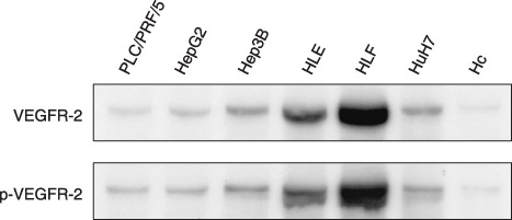
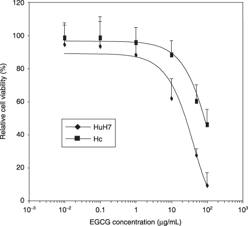
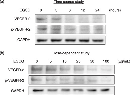
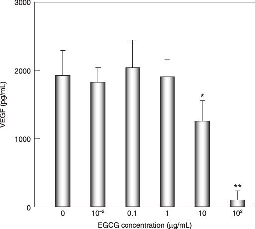
The receptor tyrosine kinase vascular endothelial growth factor (VEGF) receptor (VEGFR) plays an important role in tumor angiogenesis of hepatocellular carcinoma (HCC). (-)-Epigallocatechin gallate (EGCG), the major biologically active component of green tea, inhibits growth in a variety of human cancer cells by inhibiting the activation of several types of receptor tyrosine kinases. In this study, we examined the effects of EGCG on the activity of the VEGF-VEGFR axis in human HCC cells. The levels of total and phosphorylated (i.e. activated) form of VEGFR-2 protein (p-VEGFR-2) were observed to increase in a series of human HCC cell lines in comparison to the Hc normal human hepatocytes. EGCG preferentially inhibited the growth of HuH7 HCC cells, which express constitutive activation of the VEGF-VEGFR axis, in comparison to Hc cells. Treatment of HuH7 cells with EGCG caused a time- and dose-dependent decrease in the expression of VEGFR-2 and p-VEGFR-2 proteins. The production of VEGF from HuH7 cells was reduced by treatment with EGCG. Drinking of EGCG significantly inhibited the growth of HuH7 xenografts in nude mice and this was associated with inhibition of the activation of VEGFR-2 and its related downstream signaling molecules, including ERK and Akt. EGCG drinking also decreased the expression of Bcl-x(L) protein and VEGF mRNA in the xenografts. These findings suggest that EGCG can exert, at least in part, its growth-inhibitive effect on HCC cells by inhibiting the VEGF-VEGFR axis. EGCG might therefore be useful in the treatment of HCC.


Figures

Figure 1
Figure 1
The expression levels of total vascular endothelial growth factor receptor (VEGFR)‐2 and phosphorylated vascular endothelial growth factor receptor (p‐VEGFR)‐2 proteins in human hepatocellular carcinoma cell lines and Hc normal hepatocytes. Total protein extracts were prepared from 70% confluent cultures of the indicated cell lines and equivalent amounts of protein (20 µg/lane) were examined by western blot analysis using appropriate antibodies. Repeat western blots gave similar results.
Figure 2
Figure 2
Inhibition of cell growth by (–)‐epigallocatechin gallate (EGCG) in HuH7 human hepatocellular carcinoma cells and Hc normal hepatocytes. These cells were treated with the indicated concentrations of EGCG or DMSO for 48 h and cell viability assays were conducted using the MTT system. Results are expressed as a percentage of growth with 100% representing control cells treated with DMSO alone. Bars, SD of triplicate assays.
Figure 3
Figure 3
Effects of (–)‐epigallocatechin gallate (EGCG) on expression levels of total vascular endothelial growth factor receptor (VEGFR)‐2 and phosphorylated vascular endothelial growth factor receptor (p‐VEGFR)‐2 proteins in HuH7. The cells were treated with (a) 25 µg/mL EGCG for the indicated times (0, 3, 6, 12, and 24 h, time course study) or (b) the indicated concentration of EGCG (0, 5, 10, 25, 50, and 100 µg/mL, dose‐dependence study) for 6 h, and the cell extracts were then examined by western blot analysis using the respective antibodies. An antibody to GAPDH served as a loading control. Similar results were obtained in a repeat experiment.
Figure 4
Figure 4
Effects of (–)‐epigallocatechin gallate (EGCG) on production of vascular endothelial growth factor (VEGF) by HuH7 cells. The cells were treated with the indicated concentration of EGCG (0, 0.01, 0.1, 1.0, 10, and 100 µg/mL) in serum‐free medium for 24 h. The medium was then collected and assayed for VEGF using an ELISA kit. Bars, SD of triplicate assays. *P < 0.05, **P < 0.01: significant differences obtained by comparison with EGCG‐untreated control group.
Figure 5
Figure 5
Effects of (–)‐epigallocatechin gallate (EGCG) on the growth of HuH7 xenografts in nude mice. Male BALB/c nude mice were injected subcutaneously with 5 × 106 HuH7 cells. One week after the injection, the mice were divided into three groups and treated with following conditions for 5 weeks: group 1, control group (tap water drinking group, formula image
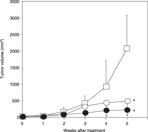
); group 2, 0.01% EGCG‐drinking group (formula image); and group 3, 0.1% EGCG‐drinking group (formula image). The growth curve of HuH7 tumors in each group are represented. Bars, SD. *P < 0.05: significant differences obtained by comparison with EGCG‐untreated control group.
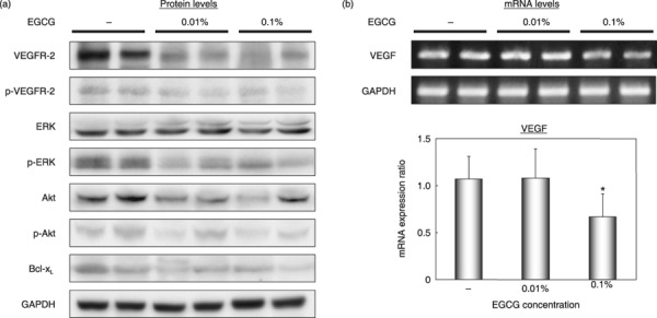
Figure 6
Figure 6
Effects of (–)‐epigallocatechin gallate (EGCG) on activation of vascular endothelial growth factor receptor (VEGFR)‐2, its related downstream signaling pathways, and on the cellular levels of Bcl‐xL proteins and vascular endothelial growth factor (VEGF) mRNA in HuH7 xenografts. The xenografts were excised from each animal at the termination of the experiment and tumor extracts were examined by (a) western blot analysis using the respective antibodies or (b) a semiquantitative RT‐PCR analysis using VEGF‐specific primers. An antibody to GAPDH served as a loading control (A). Amplified PCR products obtained with GAPDH‐specific primers served as internal controls. (b) The results obtained from RT‐PCR analysis were quantified by densitometry and are displayed in the lower panel. Bars, SD of triplicate assays. *P < 0.05: significant differences obtained by comparison with EGCG‐untreated control group. p‐, phosphorylated.

References

    1. El‐Serag HB, Rudolph KL. Hepatocellular carcinoma: epidemiology and molecular carcinogenesis. Gastroenterology 2007; 132: 2557–76. - PubMed
    1. Llovet JM, Bruix J. Molecular targeted therapies in hepatocellular carcinoma. Hepatology 2008; 48: 1312–27. - PMC - PubMed
    1. Pang RW, Poon RT. From molecular biology to targeted therapies for hepatocellular carcinoma: the future is now. Oncology 2007; 72(Suppl 1): 30–44. - PubMed
    1. Tanaka S, Arii S. Molecularly targeted therapy for hepatocellular carcinoma. Cancer Sci 2009; 100: 1–8. - PMC - PubMed
    1. Llovet JM, Ricci S, Mazzaferro V et al . Sorafenib in advanced hepatocellular carcinoma. N Engl J Med 2008; 359: 378–90. - PubMed

Publication types

MeSH terms

Substances