. 2009 Jul;85(1):40-52.
doi: 10.1016/j.ajhg.2009.06.004. Epub 2009 Jun 25.

Mutation in the AP4M1 gene provides a model for neuroaxonal injury in cerebral palsy

Affiliations

Mutation in the AP4M1 gene provides a model for neuroaxonal injury in cerebral palsy

Annemieke J M H Verkerk et al. Am J Hum Genet. 2009 Jul.

Abstract

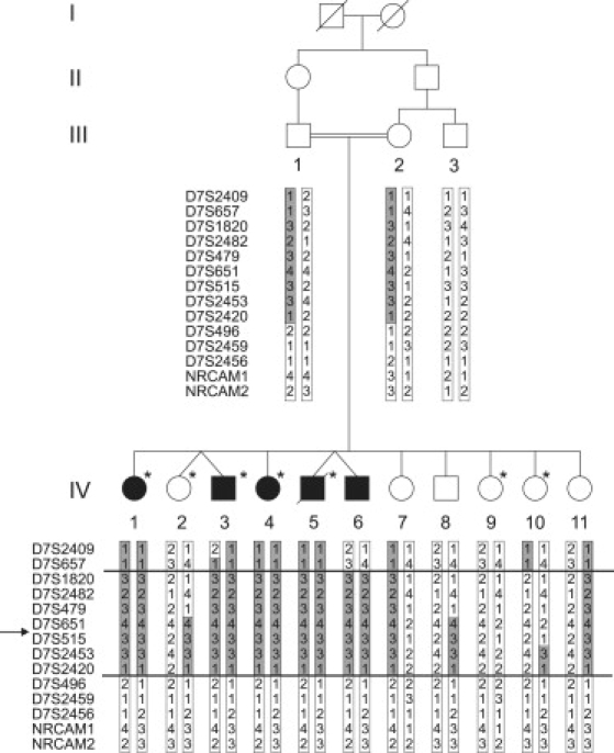
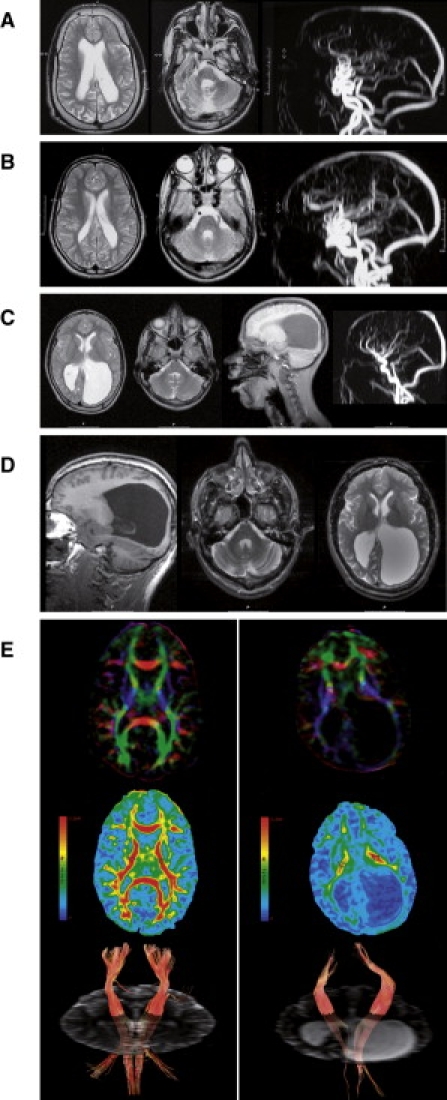
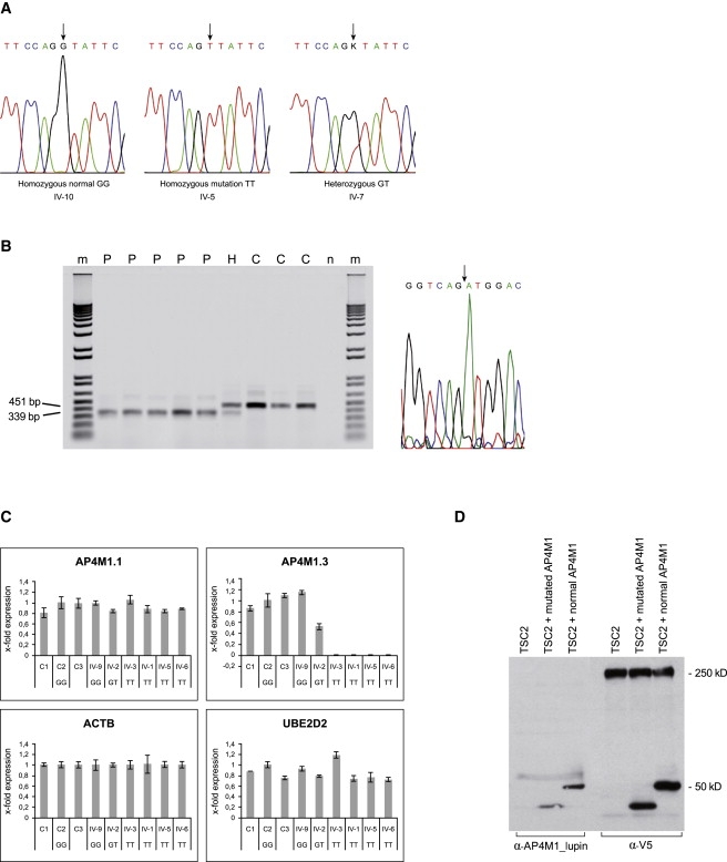
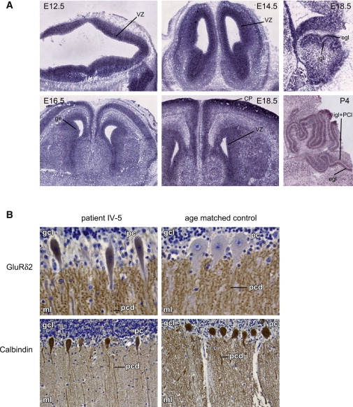
Cerebral palsy due to perinatal injury to cerebral white matter is usually not caused by genetic mutations, but by ischemia and/or inflammation. Here, we describe an autosomal-recessive type of tetraplegic cerebral palsy with mental retardation, reduction of cerebral white matter, and atrophy of the cerebellum in an inbred sibship. The phenotype was recorded and evolution followed for over 20 years. Brain lesions were studied by diffusion tensor MR tractography. Homozygosity mapping with SNPs was performed for identification of the chromosomal locus for the disease. In the 14 Mb candidate region on chromosome 7q22, RNA expression profiling was used for selecting among the 203 genes in the area. In postmortem brain tissue available from one patient, histology and immunohistochemistry were performed. Disease course and imaging were mostly reminiscent of hypoxic-ischemic tetraplegic cerebral palsy, with neuroaxonal degeneration and white matter loss. In all five patients, a donor splice site pathogenic mutation in intron 14 of the AP4M1 gene (c.1137+1G-->T), was identified. AP4M1, encoding for the mu subunit of the adaptor protein complex-4, is involved in intracellular trafficking of glutamate receptors. Aberrant GluRdelta2 glutamate receptor localization and dendritic spine morphology were observed in the postmortem brain specimen. This disease entity, which we refer to as congenital spastic tetraplegia (CST), is therefore a genetic model for congenital cerebral palsy with evidence for neuroaxonal damage and glutamate receptor abnormality, mimicking perinatally acquired hypoxic-ischemic white matter injury.


Figures

Figure 1
Figure 1
Pedigree and Haplotypes Linkage Region Pedigree of the family with four generations (I–IV). Five siblings in generation IV (IV-1, IV-3, IV-4, IV-5, and IV-6) are affected by CST (filled symbols). Empty symbols represent those unaffected for CST; the asterisk represents those affected with arterial tortuosity syndrome. Haplotypes with chromosome 7 microsatellite markers are indicated. The assumed affected haplotype is indicated in gray. Recombination events delineating the region of homozygosity in the probands are indicated with a horizontal line. The proximal boundary is determined by a recombination event in individual IV-6 between D7S657 and D7S1820. A historical recombination event that was already present in both parents between markers D7S2420 and D7S496 delimits the distal border. “→” indicates the location of the AP4M1 gene.
Figure 2
Figure 2
Brain Magnetic Resonance Imaging and DTI (A) T2 weighted brain MRI of patient IV-3 at the age of 15 years shows enlarged and asymmetric lateral ventricles, thin corpus callosum, white matter loss, with normal signal intensity, thickened skull (left), diffuse cerebellar atrophy, wide IVth ventricle, and hypoplastic pons and vermis (center). Magnetic resonance angiography (MRA) shows intra- and extracranial tortuosity of the large vessels (right), in light of the fact that this proband is also affected by ATS. (B) T2 weighted brain MRI of patient IV-4 at the age of 14 years shows enlarged lateral ventricles, particularly in the occipital area, with irregular contour and white matter loss with normal intensity and wide IVth ventricle (left and center). MRA shows arterial tortuosity of the neck and intracerebral vessels, related to ATS (right). (C) Conventional MRI of patient IV-6, performed at the age of 13 years, shows on T2 weighted images (first and second from left) diffuse but asymmetric white matter loss with massive enlargement of the occipital horns. Stretched gyri and asymmetry of the thalami suggest hydrocephalus in the past (first left and second from right). Signal intensity of the residual white matter is normal. No vascular tortuosity at the angiography (right), conform to heterozygosity for ATS. Mild cerebellar atrophy is present in T1 sagittal images (second from right). (D) Repeat MRI at age 21 years of patient IV-6 showing similar abnormalities with only minimal progression of cerebellar atrophy. (E) DTI brain images of the patient (right panel) and age- and sex-matched control (left panel). Top images show color maps: color maps are based on major eigenvector orientation in each of the voxels with red representing right to left, green antero-posterior, and blue supero-inferior anatomical directions. Middle images show fractional anisotropy maps with an example of regions of interest (ROIs, round-shaped, 30 pixels) placed in the posterior limb of the internal capsule (PLIC) (circles). For isotropic diffusion, FA is zero (no anisotropy = blue). If there is a strongly preferred direction of diffusion (indicating capture of white matter tracts in the ROI), FA approaches a value of 1 (red). Bottom images show fiber track images of the corticospinal tract with ROIs as seedpoints (minimum anisotropy threshold 0.18). Mapping the directional principal eigenvectors forms the basis for tractography with the assumption that the principal eigenvector is aligned with the direction of the fiber bundle.
Figure 3
Figure 3
Sequencing and Functional Analysis (A) Sequence analysis showing the AP4M1 mutation in intron 14 (c.1137+1G→T). The normal sequence from exon 14 to intron 14 is TTCCAGgtattc, with the last six bases of exon 14 indicated in capital letters and the first six bases of intron 14 in lowercase letters. Homozygous normal sequence, homozygous mutation as seen in probands IV-1, -3, -4, -5, and -6, and heterozygous mutation in carriers (parents III-1,2; sibs IV-2, -7, -8, and -11) are indicated by an arrow. (B) The left panel shows RT-PCR on fibroblast RNA of patients IV-1, -3, -4, -5, and -6 (P), controls (C), and a heterozygous carrier (H). Normal product length of 451 bp and reduced product length of 339 bp are indicated. n = negative control without DNA, m = 1 kb plus marker (Invitrogen). In the right panel is the RT-PCR sequence showing the exon 13 to exon 15 junction fragment, GGTCAGATGGAC, present in the patients. The arrow indicates the transition from exon 13 to exon 15. Sequence data from patient IV-4 is shown. All patients showed the same sequence. (C) qRT-PCR, expression of AP4M1 in fibroblast RNA of control samples (C1-C3, unrelated, plus family member IV-9), patients (IV-1, -3,-5, and -6), and one heterozygote (IV-2). Values are expressed as x-fold expression relative to expression of the housekeeping gene ACTB, which is arbitrarily set to 1. Expression of the housekeeping gene UBE2D2 relative to ACTB is used as positive control. Error bars indicate the standard deviation (n = 3). Expression of UBE2D2 and AP4M1.1 (primers spanning exon 2 and 3) were comparable in all control samples and patients. AP4M1.3 primers (spanning exon 14–15) showed normal mRNA expression in all control samples, 0.5-fold expression in the heterozygous family member (IV-2), and a lack of expression on all patients tested. The following abbreviations are used: GG, normal sequence; GT, heterozygous carrier; and TT, patient with homozygous mutation. (D) Immunoblot analysis of V5-tagged wild-type and mutated AP4M1 expressed in HEK293 cells, transfected with the wild-type, and mutated AP4M1 cDNA. α-V5 as well as α-AP4M1_lupin antibodies visualized an expected protein band of 50 kDa from the wild-type cDNA construct and a band of reduced size from the mutated construct. V5-tagged cDNA constructs of TSC2 gene (250 kDa) were transfected separately and in combination with AP4M1 so that the system for transfection efficiency could be tested.
Figure 4
Figure 4
mRNA In Situ Hybridization and Immunohistochemistry (A) mRNA ish expression patterns of Ap4m1 in mouse brain of different developmental stages (E, embryonal day; p, postnatal day). Ap4m1 expression is present in the ventricular zone of all four brain ventricles on days E12.5, E14.5, E16.5, and E18.5 (coronal sections, left and center panels), with expression in the ventricular zone of the lateral ventricles more sharply defined. On E16.5, pronounced expression in the upper layer of the cortical plate is present as well, broadening at E18.5 and P4. Also, expression is present in de lateral and medial ganglionic eminences (E16.5). Expression in the developing cerebellum was observed at E18.5 and P4 in the external granular and Purkinje cell layer and at P4 also in the internal granular layer (E18.5 and P4, right panel, both images showing sagittal sections). Control sense probe experiments were negative (data not shown). The following abbreviations are used: vz, ventricular zone; cp, cortical plate; ge, ganglionic eminence; egl, external granular layer; igl, internal granular layer; and pcl, Purkinje cell layer. (B) Immunohistochemical staining on brain material from patient IV-5 (17 months) (left) and an age-matched control (18 months) (right). In the upper panel, a patient's cerebellum stained with GluRδ2 revealed signal in the perikarya of the PC cells and slightly abnormally ovoid shaped perikarya as compared to the age-matched control. In both patient and controls, punctuate signals are clearly seen in the molecular layer of the cerebellum, reflecting synapse-associated signaling in dendritic spines. The punctuated GluRδ2 pattern is more pronounced in the patient compared to the control. In the lower panel, staining with PC-specific calbindin antibodies shows decreased dendritic arborization in the patient as compared to the control. The following abbreviations are used: gcl, granular cell layer; ml, molecular layer; pc, Purkinje cell body; and pcd, Purkinje cell dendrites.

References

    1. Rennie J.M., Hagmann C.F., Robertson N.J. Outcome after intrapartum hypoxic ischaemia at term. Semin. Fetal Neonatal Med. 2007;12:398–407. - PubMed
    1. Nelson K.B. Causative factors in cerebral palsy. Clin. Obstet. Gynecol. 2008;51:749–762. - PubMed
    1. Bax M., Tydeman C., Flodmark O. Clinical and MRI correlates of cerebral palsy: The European Cerebral Palsy Study. JAMA. 2006;296:1602–1608. - PubMed
    1. Volpe J.J. Neurobiology of periventricular leukomalacia in the premature infant. Pediatr. Res. 2001;50:553–562. - PubMed
    1. Folkerth R.D. Neuropathologic substrate of cerebral palsy. J. Child Neurol. 2005;20:940–949. - PubMed

Substances

Associated data