. 2009 Nov;220(1):9-22.
doi: 10.1016/j.expneurol.2009.06.012. Epub 2009 Jun 25.

Bilateral cervical contusion spinal cord injury in rats

Affiliations

Bilateral cervical contusion spinal cord injury in rats

Kim D Anderson et al. Exp Neurol. 2009 Nov.

Abstract

There is increasing motivation to develop clinically relevant experimental models for cervical SCI in rodents and techniques to assess deficits in forelimb function. Here we describe a bilateral cervical contusion model in rats. Female Sprague-Dawley rats received mild or moderate cervical contusion injuries (using the Infinite Horizons device) at C5, C6, or C7/8. Forelimb motor function was assessed using a grip strength meter (GSM); sensory function was assessed by the von Frey hair test; the integrity of the corticospinal tract (CST) was assessed by biotinylated dextran amine (BDA) tract tracing. Mild contusions caused primarily dorsal column (DC) and gray matter (GM) damage while moderate contusions produced additional damage to lateral and ventral tissue. Forelimb and hindlimb function was severely impaired immediately post-injury, but all rats regained the ability to use their hindlimbs for locomotion. Gripping ability was abolished immediately after injury but recovered partially, depending upon the spinal level and severity of the injury. Rats exhibited a loss of sensation in both fore- and hindlimbs that partially recovered, and did not exhibit allodynia. Tract tracing revealed that the main contingent of CST axons in the DC was completely interrupted in all but one animal whereas the dorsolateral CST (dlCST) was partially spared, and dlCST axons gave rise to axons that arborized in the GM caudal to the injury. Our data demonstrate that rats can survive significant bilateral cervical contusion injuries at or below C5 and that forepaw gripping function recovers after mild injuries even when the main component of CST axons in the dorsal column is completely interrupted.


Figures

Figure 1
Figure 1
Body weight temporarily decreases following cervical contusions, but returns to normal values within two weeks post-injury. (Error bars represent standard deviation; *p<0.05).
Figure 2
Figure 2
Lack of gross bladder dysfunction following cervical contusions. Bladders were expressed 3 times per day and the amount of urine expressed each morning was collected and weighed. Urine retention was used as a measure of gross bladder dysfunction. Error bars represent standard deviation.
Figure 3
Figure 3
Contusions do not produce hypersensitivity to mechanical sensory stimulation. Graphs represent the mean (+/- 95% confidence intervals) inverse response threshold of Von Frey hair stimulation to the forepaws and hindpaws of mildly (A) and moderately (B) contused rats. All animals are initially insensate to the stimuli or unable to initiate a withdrawal due to motor impairment. Sensation and/or the ability to withdraw the forepaws improves with time post-injury, as opposed to the hindpaws (*p<0.05; **p<0.001). The extent of recovered sensation is related to the severity of injury.
Figure 4
Figure 4
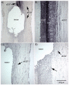
Representative tissue damage resulting from mild or moderate contusions at C7 and calculation of spared lateral tissue. A-C, horizontal sections through a moderately contused spinal cord. D-F, horizontal sections through a mildly contused spinal cord. Sections through the dorsal column are shown in panels A and D; sections through the central canal (cc) are shown in panels B and E; and sections through the ventral column are shown in panels C and F. Percent of spared lateral tissue was calculated as follows: (c+d)/[(a+b)/2] × 100 = % spared, where (a+b)/2 = control tissue and (c+d) = spared lateral tissue. Scale bar represents 1 mm for all panels.
Figure 5
Figure 5
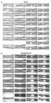
Distribution of tissue damage across the entire dorso-ventral axis of the spinal cord. A, mild contusion at C7. B, moderate contusion at C7. All sections are in the horizontal plane with rostral towards the right and caudal towards the left. Scale bar represents 1 mm for all panels. Starting in the upper left corner, sections progress down each column from ventral to dorsal.
Figure 6
Figure 6
Contusion-induced corticospinal tract damage. A, mild and moderate contusions produce complete disruption of the labeled dCST caudal to the lesion. B, the dCST and dlCST retain heavy BDA-labeling and gray matter arborization rostral to the lesion. C, spared BDA-labeled dlCST axons bypass the lesion in spared lateral white matter. D, spared BDA-labeled dlCST axons (right arrow) arborize into the gray matter caudal to the lesion (lower arrow). Scale bar represents 500 μm for B-D.
Figure 7
Figure 7
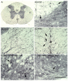
Corticospinal tract labeling caudal to the lesion. A, low magnification image of the spinal cord >2mm caudal to the lesion. Notice the absence of BDA-labeled dCST axons in the dorsal column (DC). LC = lateral column; VC = ventral column. B, high magnification image of the DC in A. Right arrow points to a rare BDA-labeled dCST axon caudal to the lesion. C, high magnification image of the LC in A. There are numerous BDA-labeled dlCST axons caudal to lesion. D, high magnification image of the VC in A. Arrows point to spared vCST axons. E, BDA-labeled dlCST arbors within the gray matter on the side ipsilateral to the labeled tract. F, BDA-labeled dlCST arbors within the gray matter on the side contralateral to the labeled tract. Scale bar represents 50 μm for B, E, andF and 100 μm for C-D.
Figure 8
Figure 8
The number of BDA-labeled axons caudal to the lesion are influenced by the severity of the contusion. For each animal group (mild n=24, moderate n=12, sham n=4), the mean number of BDA-labeled axons counted for each component of the CST (dCST, dlCST, vCST) are represented. Error bars represent standard deviation.
Figure 9
Figure 9
Grip strength recovery. A, grip strength in all rats with a mild contusion. B, gripping ability in sham-injured rats is not impaired C, grip strength in rats with a mild contusion at C6. D, grip strength in rats with a moderate contusion at C6. E, grip strength in rats with a mild contusion at the C7/8 interface. F, grip strength in rats with a moderate contusion at the C7/8 interface. Error bars represent standard error of the mean.
Figure 10
Figure 10
Grip strength recovery is influenced by the force of the contusion and the percent of lateral spared tissue at the epicenter. A, scatterplot showing the negative correlation between contusion force and mean grip strength at the end of the experiment. B, scatterplot showing the positive correlation between the percent of spared lateral tissue at the lesion epicenter and mean grip strength at the end of the experiment.

References

    1. Anderson KD. Targeting recovery: priorities of the SCI population. J. Neurotrauma. 2004;21:1371–1383. - PubMed
    1. Anderson KD, Gunawan A, Steward O. Quantitative behavioral analysis of forepaw function after cervical spinal cord injury in rats: Relationship to the corticospinal tract. Exp. Neurol. 2005;194:161–174. - PubMed
    1. Anderson KD, Gunawan A, Steward O. Spinal pathways involved in the control of forelimb motor function in rats. Exp. Neurol. 2007;206:318–331. - PubMed
    1. Anderson KD, Sharp KG, Hofstadter M, Irvine K, Murray M, Steward O. Forelimb Locomotor Assessment Scale (FLAS): Novel Assessment of Forelimb Dysfunction After Cervical SCI. Exp. Neurol. submitted. - PMC - PubMed
    1. Bareyre FM, Kerschensteiner M, Raineteau O, Mettenleiter TC, Weinmann O, Schwab ME. The injured spinal cord spontaneously forms a new intraspinal circuit in adult rats. Nat. Neurosci. 2004;7:269–277. - PubMed

Publication types

MeSH terms