

Dermoid cysts presenting as enlarged thyroid glands in a cat
- PMID: 19560384
- PMCID: PMC11132571
- DOI: 10.1016/j.jfms.2009.02.005
Dermoid cysts presenting as enlarged thyroid glands in a cat
Abstract
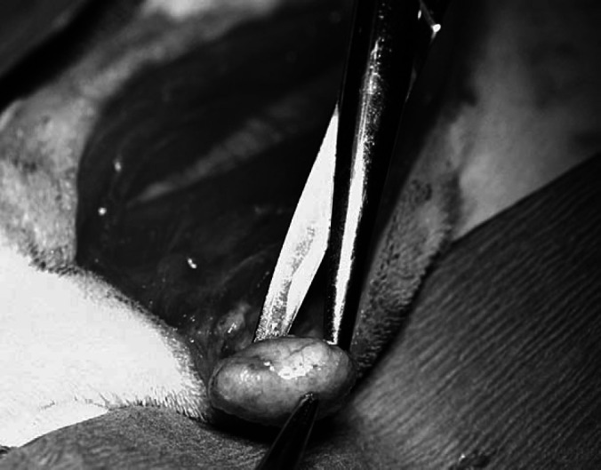
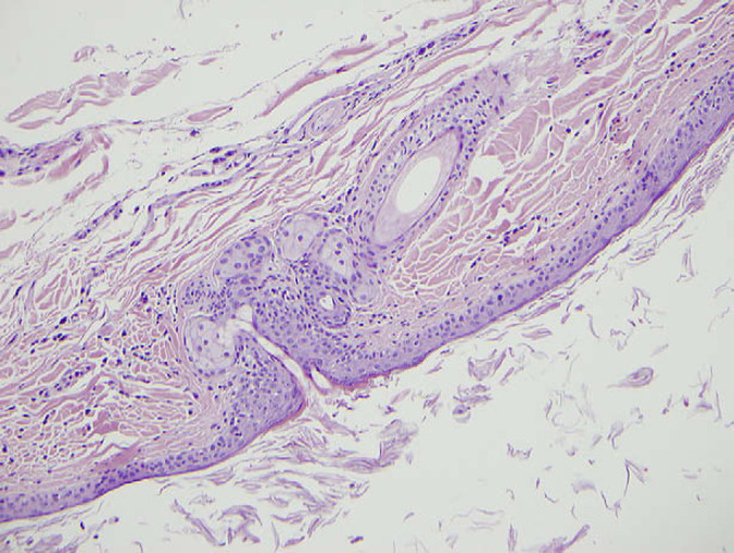
A 5-year-old spayed female cat was evaluated for hyperthyroidism based on an elevated free thyroxine (T(4)) measurement and bilaterally enlarged symmetric subcutaneous masses in the area of the thyroid glands. Physical examination revealed bilateral subcutaneous masses on either side of the cervical trachea. Blood was obtained for serum biochemical profile and thyroid function analysis. Mild hyperalbuminemia, mild hypercalcemia, and mildly increased alanine aminotransferase activity were identified. Serum concentrations of total and free thyroxine were within the reference interval. Cytologic analysis of fine-needle aspirates from one of the masses was suspicious for neoplasia. Nuclear scintigraphy revealed no abnormalities. Surgically obtained excisional biopsies of both masses were submitted for histopathology and diagnosed as bilateral dermoid cysts. After excisional biopsy, the patient recovered without incident. The histopathologic diagnosis of completely excised bilateral dermoid cysts indicated that no further medical or surgical intervention was required. This is the first report of a cat presenting with bilateral dermoid cysts in the area of the thyroid glands. Histopathologic examination was necessary to make a definitive diagnosis. Practitioners should include cysts in their list of differential diagnoses for ventral neck masses in cats.
Figures
Similar articles
-
Cervical dermoid sinus in a cat: case presentation and review of the literature.J Feline Med Surg. 2011 Dec;13(12):992-6. doi: 10.1016/j.jfms.2011.08.003. Epub 2011 Nov 10. J Feline Med Surg. 2011. PMID: 22079342 Free PMC article. Review.
-
Intrathyroid dermoid cyst presenting as a unilateral "Cold" nodule.Pediatr Surg Int. 2005 Sep;21(9):761-3. doi: 10.1007/s00383-005-1492-8. Epub 2005 Oct 20. Pediatr Surg Int. 2005. PMID: 16151821
-
Functional cystic thyroid adenoma in a cat.J Am Vet Med Assoc. 2001 Jul 15;219(2):190-3. doi: 10.2460/javma.2001.219.190. J Am Vet Med Assoc. 2001. PMID: 11469573
-
Case report: Spinal dermoid sinus in a Burmese cat with paraparesis.Aust Vet J. 2009 Nov;87(11):450-4. doi: 10.1111/j.1751-0813.2009.00487.x. Aust Vet J. 2009. PMID: 19857239
-
Diagnostic tests for hyperthyroidism in cats.Clin Tech Small Anim Pract. 2006 Feb;21(1):2-9. doi: 10.1053/j.ctsap.2005.12.001. Clin Tech Small Anim Pract. 2006. PMID: 16584024 Review.
Cited by
-
Pharyngeal dermoid cyst causing partial upper airway obstruction in a cat.JFMS Open Rep. 2022 Sep 28;8(2):20551169221122853. doi: 10.1177/20551169221122853. eCollection 2022 Jul-Dec. JFMS Open Rep. 2022. PMID: 36186252 Free PMC article.
-
Dermoid cyst in a domestic shorthair cat.Asian Pac J Trop Biomed. 2012 Mar;2(3):247-9. doi: 10.1016/S2221-1691(12)60051-3. Asian Pac J Trop Biomed. 2012. PMID: 23569907 Free PMC article.
-
Primary hyperparathyroidism due to a cystic parathyroid adenoma in a cat.Open Vet J. 2019 Jul;9(2):109-113. doi: 10.4314/ovj.v9i2.3. Epub 2019 Apr 8. Open Vet J. 2019. PMID: 31360648 Free PMC article.
References
-
- Som P.A., Sacher M., Lanzieri C.F., et al. Parenchymal cysts of the lower neck, Radiology 157, 1985, 399–406. - PubMed
-
- Baker K.P., Thomsett L.R. Canine and feline dermatology, 1990, Blackwell Scientific Publications: Cambridge, Massachusetts.
-
- Gross T., Ihrke P., Walder E., Affolter V. Skin diseases of the dog and cat, clinical and histologic diagnosis, 2005, Blackwell Publishing: Denmark.
-
- Görür K., Talas D., Özcan C. An unusual presentation of neck dermoid cyst, Eur Arch Otorhinolaryngol 262, 2005, 353–355. - PubMed
-
- Gross T., Ihrke P., Walder E. Veterinary dermatopathology, 1992, Mosby Publishing: St Louis.
Publication types
MeSH terms
LinkOut - more resources
Full Text Sources
Medical
Miscellaneous
