Review
. 2009 Dec;30(6):423-30.
doi: 10.1016/j.mam.2009.06.001. Epub 2009 Jun 26.

The adenylyl cyclase activity of anthrax edema factor

Affiliations
Review

The adenylyl cyclase activity of anthrax edema factor

Wei-Jen Tang et al. Mol Aspects Med. 2009 Dec.

Abstract

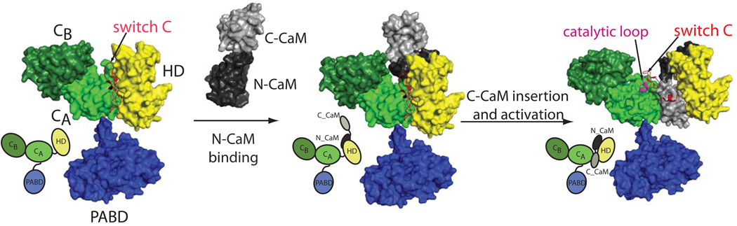
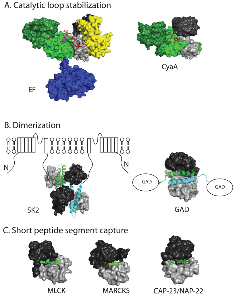
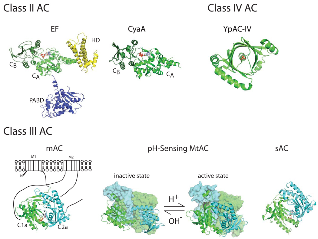
Bacillus anthracis, the etiologic agent for anthrax, secretes edema factor (EF) to disrupt intracellular signaling pathways. Upon translocation into host cells and association with a calcium sensor, calmodulin (CaM), EF becomes a highly active adenylyl cyclase (AC) that raises the intracellular concentration of cyclic AMP (cAMP). Growing evidence shows that EF plays a key role in anthrax pathogenesis by affecting cellular functions vital for host defense. This strategy is also used by Bordetella pertussis, a bacterium that causes whooping cough. Pertussis bacteria secrete the bifunctional toxin CyaA which raises the intracellular cAMP. Here, we discuss recent advances from structural analyses that reveal the molecular basis of the conserved mechanism of activation and catalysis of EF and CyaA by CaM even though these two toxins use the completely different sequences to bind CaM. Comparison of the biochemical and structural characteristics of these two AC toxins with host ACs reveal that they have diverse strategies of catalytic activation, yet use the same two-metal-ion catalytic mechanism.


Figures

Figure 1
Figure 1. Model for the catalytic activation of EF by CaM
Catalytic core domains, CA and CB, helical domain (HD) and PA binding domain (PABD) of EF are colored in light green, dark green, yellow, and purple, respectively. The N- and C-terminal domains of CaM are colored black and grey, respectively. The key switch region and the activation catalytic loop are colored in red and magenta, respectively. EF alone structure is a model based on the AC domain of EF (pdb code 1k8t) with the insertion of the PABD domain from the CaM-bound EF structure (pdb code 1xfv) (Drum et al. 2002; Shen et al. 2005). The transition state for the initial contact of N-CaM to the helical domain of EF is modeled based on the NMR study (Ulmer et al. 2003). For better visualization of the domain organization, the cartoons depicting these three states are shown in the lower left panel.
Figure 2
Figure 2. Three modes of CaM binding to its effectors
The PDB accession codes for CaM bound anthrax EF (EF), C-CaM bound pertussis CyaA (CyaA), CaM bound rat small conductance potassium channel (SK2), CaM-bound C-terminal end of glutamic acid decarboxylase (GAD), and CaM-bound fragments of myosin light chain kinase (MLCK), myristoylated alanine-rich C-kinase substrate (MARCKS), and CAP-23/NAP-22 are 1xfv, 1yrt, 1g4y, 1nwd, 1cdl, 1iwq, and 1l7z, respectively. The N- and C-terminal CaM are colored black and grey, respectively and the color scheme of EF is same as figures 1. The domains in CyaA is colored according to those of EF. Specifically, CA and CB domain of CyaA is colored as light and dark green, respectively. The key switch region and the activation catalytic loop are colored in red and magenta, respectively. The N-terminal CaM is modeled in based on the biochemical analysis and computational simulation as described (Guo et al. 2008). The CaM binding segments that are involved in the dimerization of SK2 and GAD are colored in green and cyan, respectively.
Figure 3
Figure 3. Structural comparisons of three classes (II–IV) of AC
The PDB accession codes for anthrax EF and pertussis CyaA of class II AC, membrane bound mammalian AC (mAC), bicarbonate activated soluble AC from cyanobacteria (sAC), and low pH activated inactive and active forms of AC from Mycobacteria tuberculosis (MtAC) of class III AC, and Yersinia pestis AC of class IV AC are 1xfv, 1zot, 1cjk, 1wc5, 1y10, 1y11, and 2fjt, respectively. The color scheme of class II AC is same as figure 2. The heterodimeric catalytic domains of mAC and homodimeric catalytic domains of sAC and MtAC are depicted as ribbons colored in cyan and green. For the clarity, the regulatory domains of MtAC are shown as the surface representation and colored according to their catalytic domains. YpAC is a dimer and only monomer is depicted since the catalytic center is at the center of the protein. The ATP analogues in EF, CyaA, mAC, and sAC as well as the phosphate in YpAC denote the catalytic site of these enzymatic and are colored according to their atoms.

References

    1. Abrami L, Reig N, van der Goot FG. Anthrax toxin: the long and winding road that leads to the kill. Trends Microbiol. 2005;13(2):72–78. - PubMed
    1. Bahler M, Rhoads A. Calmodulin signaling via the IQ motif. FEBS Lett. 2002;513(1):107–113. - PubMed
    1. Baldari CT, Tonello F, Paccani SR, Montecucco C. Anthrax toxins: A paradigm of bacterial immune suppression. Trends lmmunol. 2006;27(9):434–440. - PubMed
    1. Benkovic SJ, Hammes-Schiffer S. A perspective on enzyme catalysis. Science. 2003;301(5637):1196–1202. - PubMed
    1. Biel M, Michalakis S. Cyclic nucleotide-gated channels. Handb Exp Pharmacol. 2009;(191):111–136. - PubMed

Publication types

MeSH terms

LinkOut - more resources