

Respiratory recovery following high cervical hemisection
- PMID: 19560562
- PMCID: PMC2783827
- DOI: 10.1016/j.resp.2009.06.014
Respiratory recovery following high cervical hemisection
Abstract
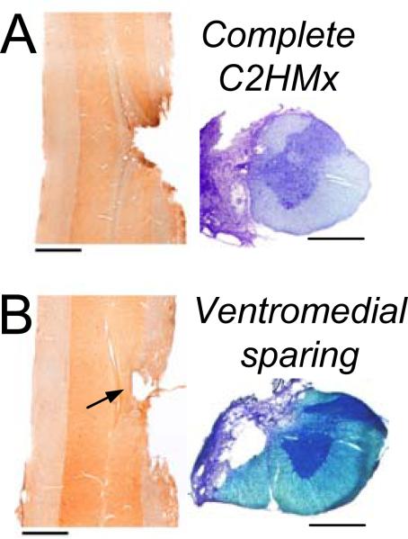
In this paper we review respiratory recovery following C2 spinal cord hemisection (C2HS) and introduce evidence for ipsilateral (IL) and contralateral (CL) phrenic motor neuron (PhrMN) synchrony post-C2HS. Rats have rapid, shallow breathing after C2HS but ventilation ( logical or (E)) is maintained. logical or (E) deficits occur during hypercapnic challenge reflecting reduced tidal volume (VT), but modest recovery occurs by 12 wks post-injury. IL PhrMN activity recovers in a time-dependent manner after C2HS, and neuroanatomical evidence suggests that this may involve both mono- and polysynaptic pathways. Accordingly, we used cross-correlation to examine IL and CL PhrMN synchrony after C2HS. Uninjured rats showed correlogram peaks consistent with synchronous activity and common synaptic input. Correlogram peaks were absent at 2 wks post-C2HS, but by 12 wks 50% of rats showed peaks occurring with a 1.1+/-0.19ms lag from zero on the abscissa. These data are consistent with prolonged conduction time to IL (vs. CL) PhrMNs and the possibility of polysynaptic inputs to IL PhrMNs after chronic C2HS.
Figures

Similar articles
-
Modest spontaneous recovery of ventilation following chronic high cervical hemisection in rats.Exp Neurol. 2008 May;211(1):97-106. doi: 10.1016/j.expneurol.2008.01.013. Epub 2008 Feb 1. Exp Neurol. 2008. PMID: 18308305 Free PMC article.
-
Graded unilateral cervical spinal cord injury and respiratory motor recovery.Respir Physiol Neurobiol. 2009 Feb 28;165(2-3):245-53. doi: 10.1016/j.resp.2008.12.010. Epub 2008 Dec 30. Respir Physiol Neurobiol. 2009. PMID: 19150658 Free PMC article.
-
Recovery of phrenic activity and ventilation after cervical spinal hemisection in rats.J Appl Physiol (1985). 2006 Mar;100(3):800-6. doi: 10.1152/japplphysiol.00960.2005. Epub 2005 Nov 3. J Appl Physiol (1985). 2006. PMID: 16269524
-
Descending bulbospinal pathways and recovery of respiratory motor function following spinal cord injury.Respir Physiol Neurobiol. 2009 Nov 30;169(2):115-22. doi: 10.1016/j.resp.2009.08.004. Epub 2009 Aug 12. Respir Physiol Neurobiol. 2009. PMID: 19682608 Review.
-
Recovery of respiratory activity after C2 hemisection (C2HS): involvement of adenosinergic mechanisms.Respir Physiol Neurobiol. 2009 Nov 30;169(2):102-14. doi: 10.1016/j.resp.2009.07.014. Epub 2009 Aug 3. Respir Physiol Neurobiol. 2009. PMID: 19651244 Free PMC article. Review.
Cited by
-
Chemogenetic inhibition of TrkB signalling reduces phrenic motor neuron survival and size.Mol Cell Neurosci. 2023 Jun;125:103847. doi: 10.1016/j.mcn.2023.103847. Epub 2023 Mar 21. Mol Cell Neurosci. 2023. PMID: 36958643 Free PMC article.
-
Targeting Spinal Interneurons for Respiratory Recovery After Spinal Cord Injury.Cells. 2025 Feb 15;14(4):288. doi: 10.3390/cells14040288. Cells. 2025. PMID: 39996760 Free PMC article. Review.
-
Respiratory function following bilateral mid-cervical contusion injury in the adult rat.Exp Neurol. 2012 May;235(1):197-210. doi: 10.1016/j.expneurol.2011.09.024. Epub 2011 Sep 21. Exp Neurol. 2012. PMID: 21963673 Free PMC article.
-
Impact of upper cervical spinal cord hemisection on diaphragm neuromotor control.J Neurophysiol. 2025 Aug 1;134(2):698-714. doi: 10.1152/jn.00279.2025. Epub 2025 Jul 28. J Neurophysiol. 2025. PMID: 40720229 Free PMC article.
-
The impact of spinal cord injury on breathing during sleep.Respir Physiol Neurobiol. 2013 Sep 15;188(3):344-54. doi: 10.1016/j.resp.2013.06.009. Epub 2013 Jun 17. Respir Physiol Neurobiol. 2013. PMID: 23791824 Free PMC article. Review.
References
-
- Bareyre FM, Kerschensteiner M, Raineteau O, Mettenleiter TC, Weinmann O, Schwab ME. The injured spinal cord spontaneously forms a new intraspinal circuit in adult rats. Nat Neurosci. 2004;7:269–277. - PubMed
Publication types
MeSH terms
Grants and funding
LinkOut - more resources
Full Text Sources
Medical
Miscellaneous
