

Regional age-related effects in the monkey brain measured with 1H magnetic resonance spectroscopy
- PMID: 19560839
- PMCID: PMC2891318
- DOI: 10.1016/j.neurobiolaging.2009.05.020
Regional age-related effects in the monkey brain measured with 1H magnetic resonance spectroscopy
Abstract
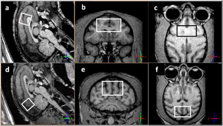
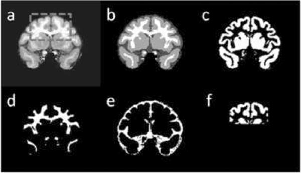
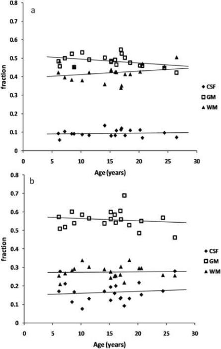
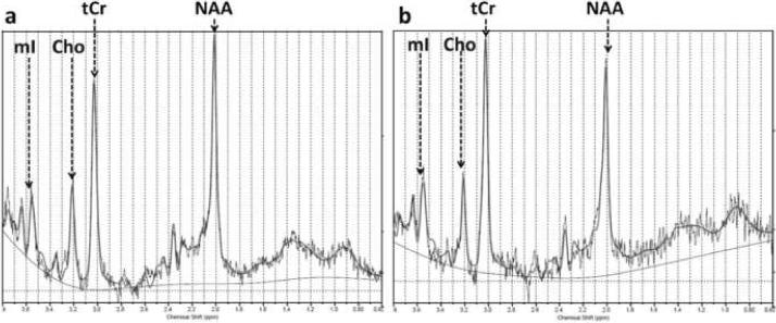
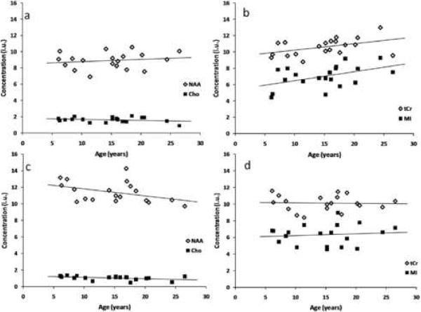
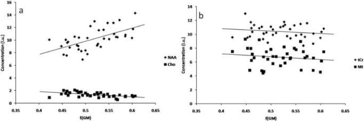
The rhesus monkey is a useful model for examining age-related effects on the brain, because of the extensive neuroanatomical homology between the monkey and the human brain, the tight control for neurological diseases as well as the possibility of obtaining relevant behavioral data and post-mortem tissue for histological analyses. Here, proton magnetic resonance spectroscopy ((1)H-MRS) was used together with high-resolution anatomical MRI images to carefully assess regional concentrations of brain metabolites in a group of 20 rhesus monkeys. In an anterior volume of interest (VOI) that covered frontal and prefrontal areas, significant positive correlations of myo-inositol and of total creatine concentrations with age were detected, whereas N-acetyl aspartate (NAA) and choline compounds (Cho) were not significantly correlated with age. In an occipito-parietal VOI, all metabolites showed no statistically significant age-dependent trend. Strong correlations were found between NAA concentration and gray matter fraction in the VOIs as well as between choline compounds and white matter fraction.
Copyright © 2009 Elsevier Inc. All rights reserved.
Figures
Similar articles
-
(1)H-magnetic resonance spectroscopy ((1)H-MRS) in methamphetamine dependence and methamphetamine induced psychosis.Schizophr Res. 2014 Mar;153(1-3):122-8. doi: 10.1016/j.schres.2014.01.029. Epub 2014 Feb 12. Schizophr Res. 2014. PMID: 24529366
-
Effects of Alzheimer disease on fronto-parietal brain N-acetyl aspartate and myo-inositol using magnetic resonance spectroscopic imaging.Alzheimer Dis Assoc Disord. 2006 Apr-Jun;20(2):77-85. doi: 10.1097/01.wad.0000213809.12553.fc. Alzheimer Dis Assoc Disord. 2006. PMID: 16772742 Free PMC article.
-
Proton magnetic resonance spectroscopy of late-life major depressive disorder.Psychiatry Res. 2009 Jun 30;172(3):210-4. doi: 10.1016/j.pscychresns.2009.01.003. Epub 2009 Mar 20. Psychiatry Res. 2009. PMID: 19303260
-
Magnetic resonance spectroscopy.J Neuroophthalmol. 2005 Sep;25(3):217-26. doi: 10.1097/01.wno.0000177307.21081.81. J Neuroophthalmol. 2005. PMID: 16148633 Review.
-
Neurochemistry of drug action: insights from proton magnetic resonance spectroscopic imaging and their relevance to addiction.Ann N Y Acad Sci. 2010 Feb;1187:148-71. doi: 10.1111/j.1749-6632.2009.05143.x. Ann N Y Acad Sci. 2010. PMID: 20201852 Free PMC article. Review.
Cited by
-
Preclinical Magnetic Resonance Imaging and Spectroscopy Studies of Memory, Aging, and Cognitive Decline.Front Aging Neurosci. 2016 Jun 29;8:158. doi: 10.3389/fnagi.2016.00158. eCollection 2016. Front Aging Neurosci. 2016. PMID: 27468264 Free PMC article. Review.
References
-
- Adalsteinsson E, Sullivan EV, Kleinhans N, Spielman DM, Pfefferbaum A. Longitudinal decline of the neuronal marker N-acetyl aspartate in Alzheimer's disease. Lancet. 2000;355(9216):1696–7. - PubMed
-
- Andersen AH, Zhang Z, Zhang M, Gash DM, Avison MJ. Age-associated changes in rhesus CNS composition identified by MRI. Brain Res. 1999;829(1-2):90–8. - PubMed
-
- Arnsten AF, Goldman-Rakic PS. Analysis of alpha-2 adrenergic agonist effects on the delayed nonmatch-to-sample performance of aged rhesus monkeys. Neurobiol Aging. 1990;11(6):583–90. - PubMed
-
- Bachevalier J, Lnandis LS, Walker LC, Brickson M, Mishkin M, Price DL, Cork LC. Aged monkeys exhibit behavioral deficits indicative of widespread cerebral dysfunction. Neurobiol Aging. 1991;12(2):99–111. - PubMed
Publication types
MeSH terms
Substances
Grants and funding
LinkOut - more resources
Full Text Sources
Other Literature Sources
Medical
