. 2009 Jul 15;183(2):1301-12.
doi: 10.4049/jimmunol.0803567. Epub 2009 Jun 26.

Autocrine IL-10 induces hallmarks of alternative activation in macrophages and suppresses antituberculosis effector mechanisms without compromising T cell immunity

Affiliations

Autocrine IL-10 induces hallmarks of alternative activation in macrophages and suppresses antituberculosis effector mechanisms without compromising T cell immunity

Tanja Schreiber et al. J Immunol. .

Abstract

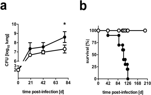
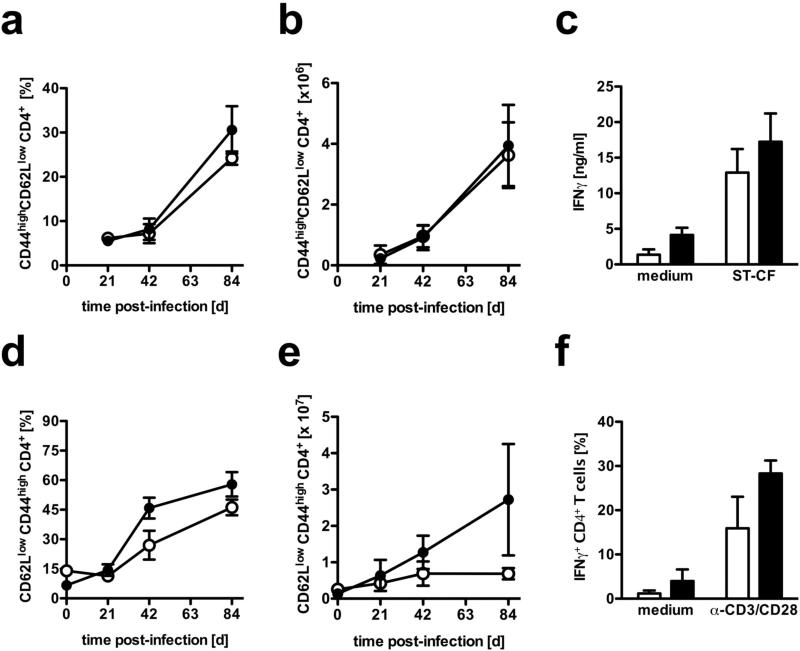
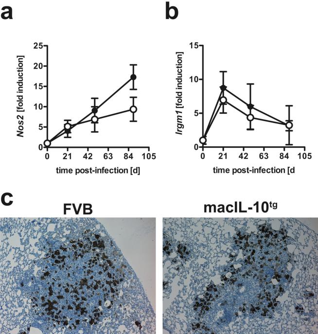
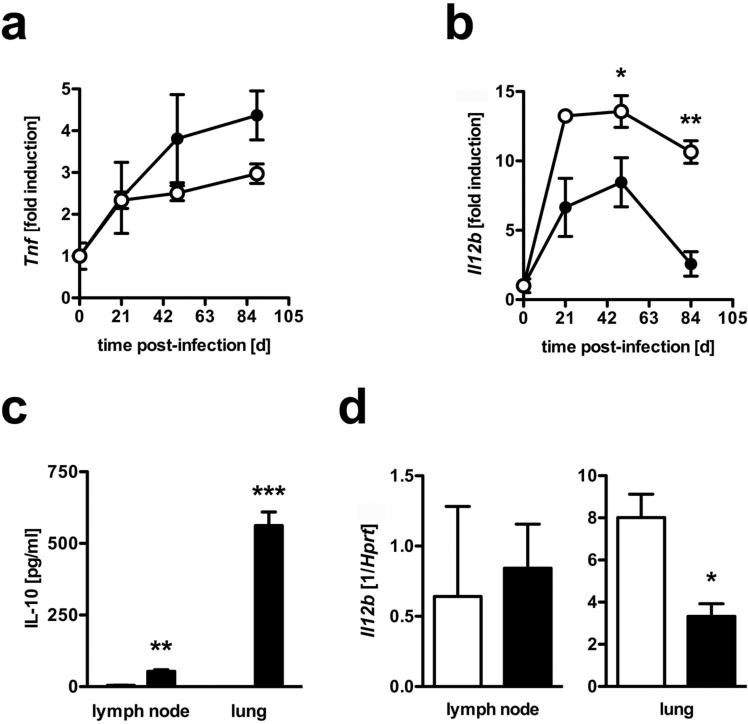
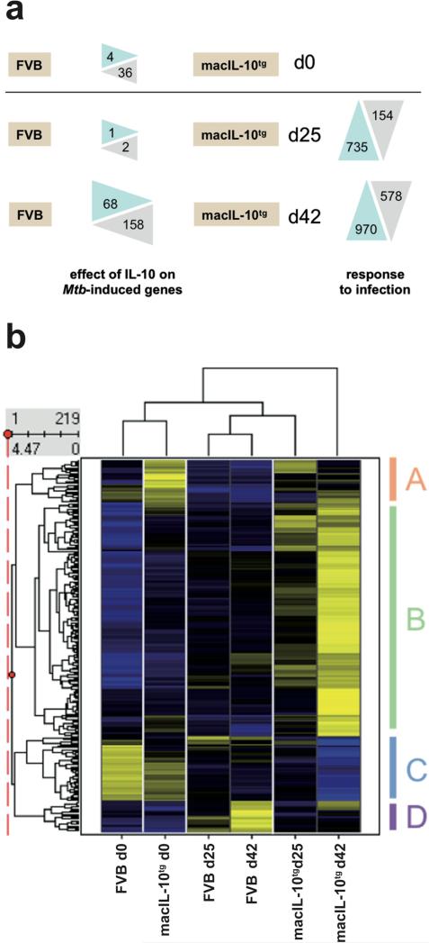
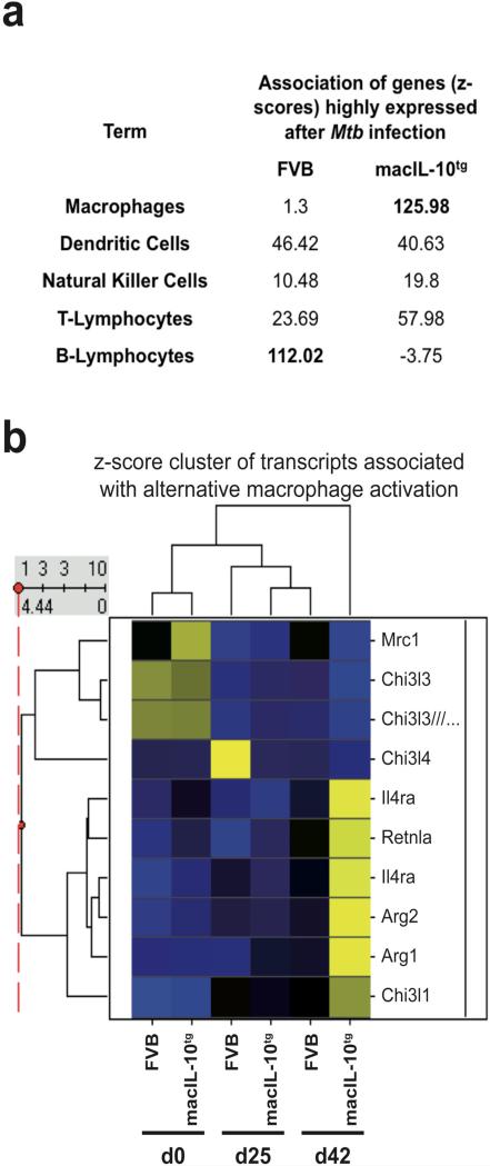
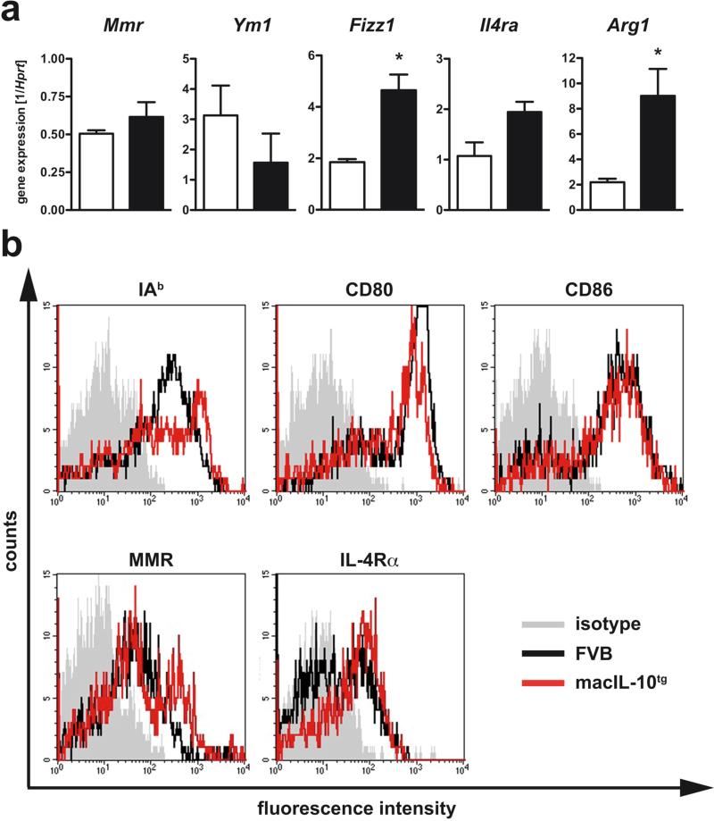
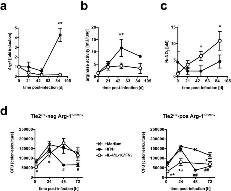
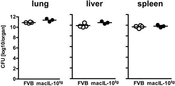
Elevated IL-10 has been implicated in reactivation tuberculosis (TB). Since macrophages rather than T cells were reported to be the major source of IL-10 in TB, we analyzed the consequences of a macrophage-specific overexpression of IL-10 in transgenic mice (macIL-10-transgenic) after aerosol infection with Mycobacterium tuberculosis (Mtb). MacIL-10 transgenic mice were more susceptible to chronic Mtb infection than nontransgenic littermates, exhibiting higher bacterial loads in the lung after 12 wk of infection and dying significantly earlier than controls. The differentiation, recruitment, and activation of Th1 cells as well as the induction of IFN-gamma-dependent effector genes against Mtb were not affected by macrophage-derived IL-10. However, microarray analysis of pulmonary gene expression revealed patterns characteristic of alternative macrophage activation that were overrepresented in Mtb-infected macIL-10 transgenic mice. Importantly, arginase-1 gene expression and activity were strikingly enhanced in transgenic mice accompanied by a reduced production of reactive nitrogen intermediates. Moreover, IL-10-dependent arginase-1 induction diminished antimycobacterial effector mechanisms in macrophages. Taken together, macrophage-derived IL-10 triggers aspects of alternative macrophage activation and promotes Mtb recrudescence independent of overt effects on anti-TB T cell immunity.


Figures

Figure 1
Figure 1. macIL-10tg mice are highly susceptible to Mtb infection
FVB (open symbols) and macIL-10tg (solid symbols) mice were infected with 100 CFU Mtb via the aerosol route. (a) For mycobacterial colony enumeration assays, lungs, were removed at the indicated time points and colony forming units were counted 21 days after plating of serial diluted organ homogenates. Data represent means and standard deviations of 4 mice. One experiment representative of three performed is shown. Statistical analysis was performed using the unpaired Student's t test defining differences between FVB and macIL-10tg mice as significant (*, p≤0.05). (b) During the course of infection survival of 10 infected mice per group was monitored. Animals that lost more than 25% of their original body weight were sacrificed. Statistical analysis of the resulting survival curve was performed using the log rank test. Differences in survival kinetics between FVB and macIL-10tg mice were highly significant (p≤0.001). Therefore, the experiment was terminated and remaining wild-type animals were euthanized in compliance with ethical guidelines for animal experimentation.
Figure 2
Figure 2. Normal recruitment and activation of T cells in macIL-10tg mice after aerosol infection with Mtb
FVB (open symbols) and macIL-10tg (solid symbols) mice were infected with 100 CFU Mtb via the aerosol route. At different time points, single cell suspensions from (a-c) draining lymph nodes and (e-f) lungs were prepared. Surface and activation markers were stained for flow cytometric analysis of activated (a, b, d, e) CD44highCD62Llow CD4+ T cells. The relative (a, d) and (b, e) absolute amount of activated CD4+ T cells was calculated. Data represent means and standard deviations of 4 mice. One experiment representative of two performed is shown. (c) Antigen-specific restimulation of CD4+ T cells from mediastinal lymph nodes was performed 21 days after infection. Enriched (>95%) CD4+ T cells were cultivated in the presence of antigen-presenting peritoneal macrophages that have been pulsed with medium or ST-CF. After 72 h, the supernatants were assayed for IFNγ by ELISA. (f) Analysis of IFNγ production by CD4+ T cells from lungs of infected mice was performed 21 days after infection. For detection of intracellular IFNγ, cells were stimulated with plate-bound anti-CD3/CD28 mAb for 4 h in the presence of GolgiPlug™. After staining for CD4, cells were fixed and permeabilized and intracellularly accumulated IFNγ was detected by a PE-labelled anti- IFNγ mAb. Data represent means of triplicate assays. Data represent means and standard deviations of 3 mice. One experiment representative of two performed is shown.
Figure 3
Figure 3. Efficient induction of IFNγ-dependent effector mechanisms in Mtb-infected macIL-10tg mice
FVB (open symbols) and macIL-10tg (solid symbols) mice were infected with 100 CFU Mtb via the aerosol route. Gene-expression of IFNγ-dependent (a) Nos2 and (b) LRG-47 (lrgm1) was determined in lung homogenates from uninfected and mice infected for 21, 49 and 89 days by RTPCR based on expression of Hprt. Data represent means and standard deviations of 3 mice. One experiment representative of two performed is shown. (c) For immunohistological detection of NOS2, 2−3 μm sections were prepared from FVB and macIL-10tg mice 42 days after aerosol infection and staining was performed with a polyclonal rabbit anti-mouse NOS2 antibody. Representative results of 4 mice per group obtained in three independent experiments are shown.
Figure 4
Figure 4. Differential production of IL-10 specifically suppresses IL-12p40 in infected tissue but not in draining lymph nodes
FVB (open symbols) and macIL-10tg (solid symbols) mice were infected with 100 CFU Mtb via the aerosol route. Gene-expression of (a) Tnf and (b) IL-12p40 (Il12b) was determined in lung homogenates from uninfected and mice infected for 21, 49 and 89 days by quantitative real time RT-PCR based on expression of Hprt. (c) After 42 days of infection, 3×10⁶ cells from single cell suspensions of mediastinal lymph nodes and perfused lungs were cultured. After 24 h, the production of IL-10 was determined by ELISA. (d) Gene-expression of IL-12p40 (Il12b) was determined in mediastinal lymph node and lung homogenates from mice infected for 42 days by RT-PCR based on expression of hprt. Data represent means and standard deviations of 3 mice. One experiment representative of two performed is shown. Statistical analysis was performed using the unpaired Student's t test defining differences between FVB and macIL-10tg mice as significant (*, p≤0.05; **, p≤0.01***, p≤0.001).
Figure 5
Figure 5. Pulmonary gene expression in wildtype and macIL-10tg mice
FVB and macIL-10tg mice were infected with 100 CFU Mtb via the aerosol route. Gene-expression analysis was performed in lung homogenates from uninfected and mice infected for 25 and 42 days. (a) Numbers of probe sets regulated by overexpression of IL-10 in Mtb-infected mice. After preselection of 4188 probe sets that were regulated across the experiment, for both time points the number of probe sets are shown that are up- or down-regulated more than three-fold in WT or macIL-10tg mice (“response to infection”). Limma statistics was used to compare WT and macIL-10tg mice. For d0, all 4188 preselected probe sets were used as the basis for comparison. At d25 and d42 after infection, the 735 and 970 genes up-regulated by infection were used to define statistically significant changes due to IL-10 overexpression. (b) Hierarchical clustering of z-score normalized average expression values of 219 probe sets significantly different (Limma q value < 0.05) between FVB and macIL-10tg. Yellow and blue colour indicates high and low relative expression, respectively.
Figure 6
Figure 6. Signs of alternative macrophage activation in the lung transcriptome of macIL-10tg mice after Mtb infection
(a) Gene lists identified in Fig. 5A as upregulated differentially in FVB and macIL-10tg mice at d42 after infection were entered into the Genomatix Bibliosphere literature-based data mining tool. Shown are the z-score values indicating the degree of co-citation of selected cell types with the gene list from both genotypes. Higher z-score values indicate stronger enrichment. (b) Changes in expression of selected genes associated with alternative macrophage activation during Mtb infection of FVB and macIL-10tg mice were visualized using hierarchical clustering of z-score normalized average expression values. Similarity between experimental conditions and patterns of individual genes is indicated by dendrograms. Gene symbols are given.
Figure 7
Figure 7. Enhanced alternative macrophage activation in lungs from Mtb-infected macIL-10tg mice
FVB (open symbols) and macIL-10tg (solid symbols) mice were infected with 100 CFU Mtb via the aerosol route. (a) Gene-expression of Mmr, Ym1, Fizz1, Il4ra, Arg1 was determined in lung homogenates from mice infected for 42 days by quantitative real time RT-PCR based on expression of hprt. Statistical analysis was performed using the unpaired Student's t test defining differences between FVB and macIL-10tg mice as significant (*, p0.05). (b) Expression of activation markers on pulmonary macrophages was assessed by flowcytometric analysis of IAq, CD80, CD86, MMR, and IL-4Rα gated on PI F4/80+ cells in single cell suspensions of perfused lungs from FVB (black line) and macIL-10tg mice (red line) mice. Representative histogram of 1 out of 3 mice per group (gray histogram, isotype control).
Figure 8
Figure 8. IL-10-dependent arginase-1 induction diminished anti-mycobacterial effector mechanisms
(a-c) FVB (open symbols) and macIL-10tg (solid symbols) mice were infected with 100 CFU Mtb via the aerosol route. (a) Gene-expression of Arg1 was determined in lung homogenates from uninfected and mice infected for 21, 49 and 89 days by quantitative real time RT-PCR based on expression of hprt. Data represent means and standard deviations of 3 mice. One experiment representative of three performed is shown. (b) At different time points, the degree of arginase activity in lungs from uninfected and infected mice was determined as urea production in homogenates of weighed pieces of lungs after addition of L-arginine. (c) During the course of infection, the RNI content in sera was measured by the Griess reaction. Statistical analysis in (a) - (c) was performed using the unpaired Student's t test defining differences between FVB and macIL-10tg mice as significant (*, p≤0.05; **, p≤0.01). (d) BMMϕ were incubated with medium (crossed lines), IFNγ (solid symbols) or IL-4/IL-10/IFNγ (open symbols) and infected with Mtb at a MOI of 0.5. Colony forming units were counted 21 days after plating of serial diluted cell lysates. Data in (d) represent means and standard deviations of BMMϕ from 3 mice. ANOVA was performed using the Dunnett Multiple Comparison test), correcting for multiple testing, that defines different error probabilities between untreated cells and macrophages that have been incubated with IFNγ (#, p≤0.05; ##, p≤0.01) or IL-4/IL-10/IFNγ (*, p≤0.05; **, p≤0.01.
Figure 9
Figure 9. macIL-10tg mice control bacterial growth after M. avium infection
FVB (open circles) and macIL-10tg (solid circles) mice mice were infected with 105 CFU M. avium via the aerosol route. For mycobacterial colony enumeration assays, lungs, spleen and liver were removed 105 days after infection and colony forming units were counted 21 days after plating of serial diluted organ homogenates. Data represent means of 2 − 4 mice.

References

    1. Kaufmann SH. Tuberculosis: back on the immunologists’ agenda. Immunity. 2006;24:351–357. - PubMed
    1. Cooper AM, Dalton DK, Stewart TA, Griffin JP, Russell DG, Orme IM. Disseminated tuberculosis in interferon gamma gene-disrupted mice. J. Exp.Med. 1993;178:2243–2247. - PMC - PubMed
    1. Flynn JL, Goldstein MM, Chan J, Triebold KJ, Pfeffer K, Lowenstein CJ, Schreiber R, Mak TW, Bloom BR. Tumor necrosis factor-alpha is required in the protective immune response against Mycobacterium tuberculosis in mice. Immunity. 1995;2:561–572. - PubMed
    1. MacMicking JD, North RJ, LaCourse R, Mudgett JS, Shah SK, Nathan CF. Identification of nitric oxide synthase as a protective locus against tuberculosis. Proc. Natl. Acad. Sci. U.S.A. 1997;94:5243–5248. - PMC - PubMed
    1. MacMicking JD, Taylor GA, McKinney JD. Immune control of tuberculosis by IFN-gamma-inducible LRG-47. Science. 2003;302:654–659. - PubMed

Publication types