. 2009 Aug 21;284(34):22633-40.
doi: 10.1074/jbc.M109.026773. Epub 2009 Jun 27.

The interaction of mitochondrial iron with manganese superoxide dismutase

Affiliations

The interaction of mitochondrial iron with manganese superoxide dismutase

Amornrat Naranuntarat et al. J Biol Chem. .

Abstract

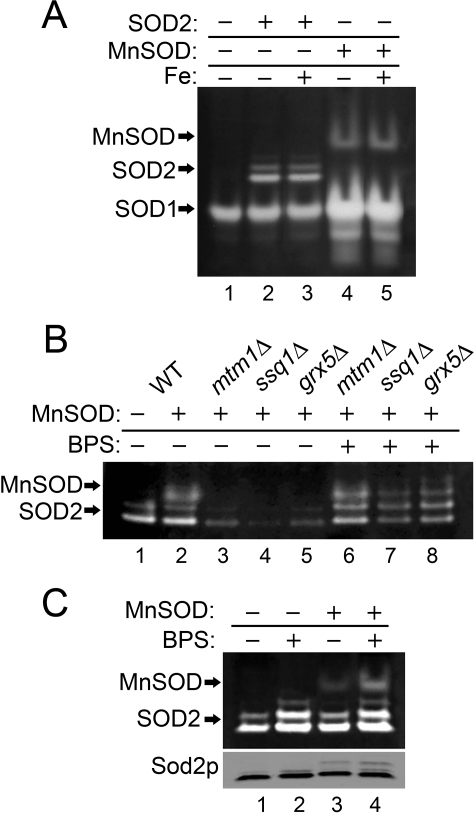
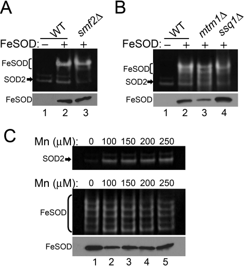
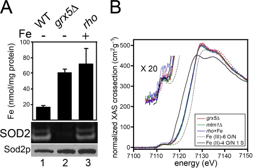
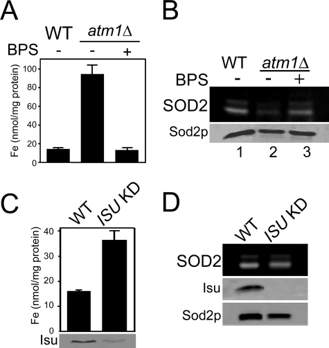
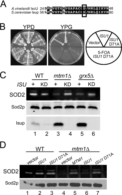
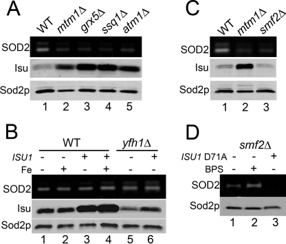
Superoxide dismutase 2 (SOD2) is one of the rare mitochondrial enzymes evolved to use manganese as a cofactor over the more abundant element iron. Although mitochondrial iron does not normally bind SOD2, iron will misincorporate into Saccharomyces cerevisiae Sod2p when cells are starved for manganese or when mitochondrial iron homeostasis is disrupted by mutations in yeast grx5, ssq1, and mtm1. We report here that such changes in mitochondrial manganese and iron similarly affect cofactor selection in a heterologously expressed Escherichia coli Mn-SOD, but not a highly homologous Fe-SOD. By x-ray absorption near edge structure and extended x-ray absorption fine structure analyses of isolated mitochondria, we find that misincorporation of iron into yeast Sod2p does not correlate with significant changes in the average oxidation state or coordination chemistry of bulk mitochondrial iron. Instead, small changes in mitochondrial iron are likely to promote iron-SOD2 interactions. Iron binds Sod2p in yeast mutants blocking late stages of iron-sulfur cluster biogenesis (grx5, ssq1, and atm1), but not in mutants defective in the upstream Isu proteins that serve as scaffolds for iron-sulfur biosynthesis. In fact, we observed a requirement for the Isu proteins in iron inactivation of yeast Sod2p. Sod2p activity was restored in mtm1 and grx5 mutants by depleting cells of Isu proteins or using a dominant negative Isu1p predicted to stabilize iron binding to Isu1p. In all cases where disruptions in iron homeostasis inactivated Sod2p, we observed an increase in mitochondrial Isu proteins. These studies indicate that the Isu proteins and the iron-sulfur pathway can donate iron to Sod2p.


Figures

FIGURE 1.
FIGURE 1.
Effects of mitochondrial iron homeostasis on a heterologous E. coli Mn-SOD. The designated strains were grown in YPD medium supplemented where indicated with 80 μm BPS or 2 mm ammonium iron(III) citrate (Fe:+). Whole-cell lysates were analyzed for SOD activity by native gel electrophoresis and nitroblue tetrazolium staining. Mn-SOD, SOD1, and SOD2 indicate positions of active Mn-SOD from E. coli and Cu/Zn-Sod1p and manganese Sod2p from S. cerevisiae, respectively. Sod2p polypeptide from the whole-cell lysates was analyzed by immunoblot (C, bottom panel). Strains utilized: the sod2Δ strain transformed where indicated with the yeast SOD2 plasmid pAN001 (A) or the E. coli Mn-SOD expression plasmid, pAN002 (B and C). The BY4741 WT (B, lanes 1 and 2, and C) or indicated mutant derivatives (B) transformed where designated (+) with plasmid pAN002 for expressing E. coli Mn-SOD.
FIGURE 2.
FIGURE 2.
Enzymatically active Fe-SOD expressed in yeast mitochondria. Strains that were transformed where indicated (Fe-SOD:+, A and B, and C, bottom panel) with the plasmid for expressing a FLAG-tagged version of E. coli Fe-SOD were analyzed for SOD activity (top) and for levels of Fe-SOD by immunoblotting with anti-FLAG (bottom). FLAG-tagged Fe-SOD typically runs as multiple bands on native gels; the major bands are shown in brackets. S. cerevisiae SOD2 activity is shown by arrows. Strains utilized: WT, BY4741; smf2Δ, 1878 (A); WT, BY4741; mtm1Δ, MY019; ssq1Δ, 5278 (B); mtm1Δ, MY019 (top panel); sod2Δ mtm1Δ, MY020 (C, bottom panel) transformed with the E. coli Fe-SOD expression plasmid.
FIGURE 3.
FIGURE 3.
Bulk mitochondrial iron is unchanged in cells accumulating SOD2-reactive iron. A, the designated strains grown where indicated in the presence of 2 mm ammonium iron(III) citrate were subjected to analysis of mitochondrial iron by atomic absorption spectrometry (values represent averages of four readings from two independent cultures, error bars indicate S.D.) (top) and to yeast Sod2 activity analysis (middle) and Sod2p protein levels (bottom) as in Fig. 1. B, normalized XANES spectra of mitochondria samples (grx5Δ, mtm1Δ, and rho supplemented with 2 mm ammonium iron(III) citrate; rho+Fe). Duplicate sample sets from independent cultures are color-coded per the inset legend, with each duplicate represented by a solid and dotted line. The expansion showing the 1s → 3d transition is enlarged 20-fold vertically. Strains utilized: WT, BY4741; grx5Δ, 2769; mtm1Δ, MY019; rho, LJ109.
FIGURE 4.
FIGURE 4.
Effects of disrupting iron-sulfur biogenesis on Sod2p activity. A and B, the designated strains were grown in YPD medium containing where indicated (BPS: +) 80 μm BPS. A, mitochondrial iron levels were measured as in 3A. B, Sod2p activity and protein levels were monitored as in Fig. 1. C and D, cells that were originally maintained on galactose-containing medium were shifted to glucose for 48 h to deplete Isu1p in the ISU knockdown (KD) cultures. C, top, mitochondrial iron levels monitored as in 3A. C, bottom, and D, whole-cell lysates were analyzed where indicated for Isu (combination of yeast Isu1p and Isu2p) protein levels by immunoblot and for Sod2p activity and protein as in Fig. 1. Strains utilized: WT, BY4741; atm1Δ, LJ206 (A and B); WT, BY4741; ISU KD, LJ403 (C and D).
FIGURE 5.
FIGURE 5.
Blocking Isu activity in yeast. A, alignment of an N-terminal segment of A. vinelandii IscU versus S. cerevisiae Isu1p. Identical and similar residues are in black and gray; boxed is a conserved aspartate targeted for mutagenesis. B, WT cells transformed with vectors for overproducing either WT Isu1p or D71A Isu1p or with empty vector were tested for growth on fermentable (YPD) and nonfermentable (YPG) carbon sources, the latter being a marker of mitochondrial respiration. Where indicated (+5-fluoroorotic acid), three independent transformants for D71A ISU1 were induced to shed the D71A ISU1 plasmid by growth on 5-fluoroorotic acid prior to growth tests. C and D, whole-cell lysates were analyzed for SOD activity (top) and Sod2p protein levels (bottom) as in Fig. 1. C, cells that were originally maintained on methionine-free medium were shifted to enriched methionine-containing YPD medium for 48 h to deplete Isu1p in the ISU KD cultures. ISU +, cells harboring wild type ISU1 and ISU2 alleles. Isup, combined levels of Isu1p and Isu2p detected by immunoblot using an anti-E. coli IscU antibody. D, WT and mtm1Δ cells were transformed with the indicated plasmids for overexpression of wild type or D71A ISU1 or for MTM1 and were grown in selecting synthetic defined medium. Strains utilized: WT, BY4741 and mtm1Δ, MY019 (A, B, and D); WT, BY4742; ISU KD, LJ436 (C); mtm1Δ, LJ443; mtm1Δ ISU KD, LJ444; grx5Δ, LJ445; grx5Δ ISU KD, LJ446.
FIGURE 6.
FIGURE 6.
Isu1p levels and iron inactivation of Sod2p. Sod2p activity and protein levels and Isu protein levels were monitored as in Fig. 5. A and C, indicated strains were grown in YPD medium. B and D, strains were grown in selecting synthetic defined medium supplemented where indicated with 2 mm ammonium iron(III) citrate (Fe:+) or 80 μm BPS. As designated (ISU1: + or ISU1 D71A: +), cells were transformed with the pLJ129 or pLJ357 vectors for overexpressing WT or D71A Isu1p. Strains utilized: WT, BY4741; mtm1Δ, MY019; grx5Δ, 2769; ssq1Δ, 5278; atm1Δ, LJ206; yfh1Δ, AN015; smf2Δ, 1878.

References

    1. Wintjens R., Noël C., May A. C., Gerbod D., Dufernez F., Capron M., Viscogliosi E., Rooman M. (2004) J. Biol. Chem. 279,9248–9254 - PubMed
    1. Carlioz A., Ludwig M. L., Stallings W. C., Fee J. A., Steinman H. M., Touati D. (1988) J. Biol. Chem. 263,1555–1562 - PubMed
    1. Whittaker J. W. (2003) Biochem. Soc. Trans. 31,1318–1321 - PubMed
    1. Vance C. K., Miller A. F. (1998) J. Am. Chem. Soc. 120,461–467
    1. Vance C. K., Miller A. F. (2001) Biochemistry 40,13079–13087 - PubMed

Publication types

MeSH terms