. 2009 Sep;32(7):677-88.
doi: 10.1097/CJI.0b013e3181ab1824.

High-avidity autoreactive CD4+ T cells induce host CTL, overcome T(regs) and mediate tumor destruction

Affiliations

High-avidity autoreactive CD4+ T cells induce host CTL, overcome T(regs) and mediate tumor destruction

Andrew G Brandmaier et al. J Immunother. 2009 Sep.

Abstract

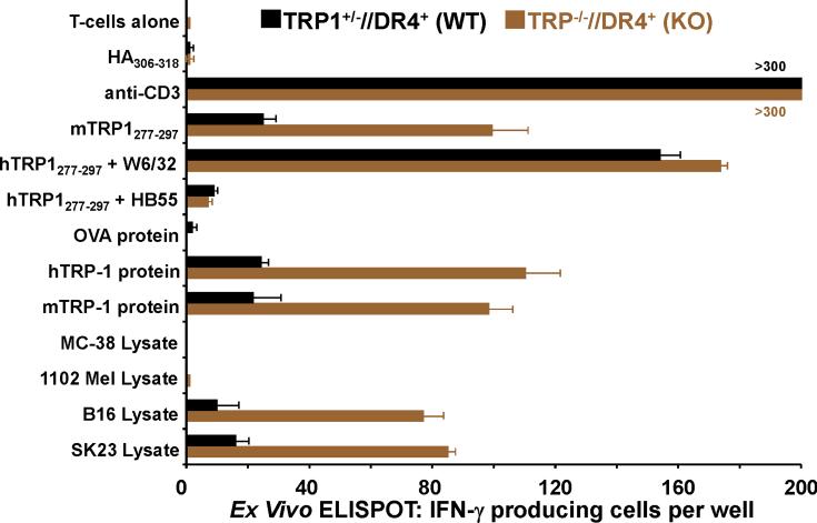
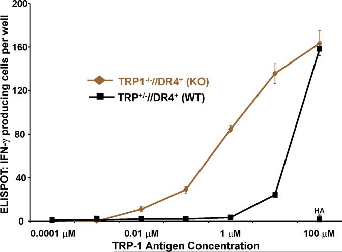
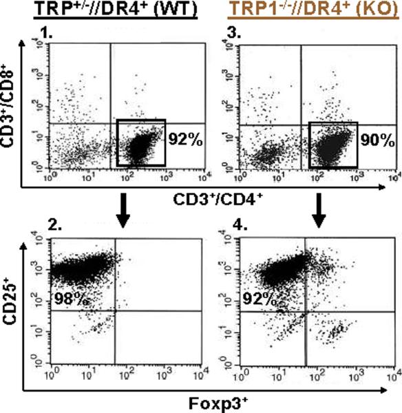
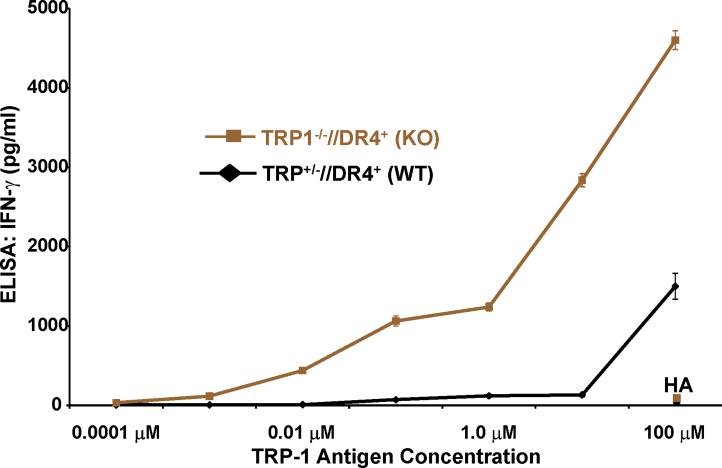
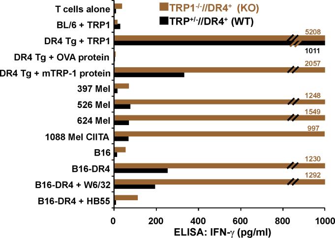
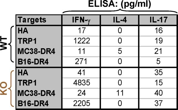
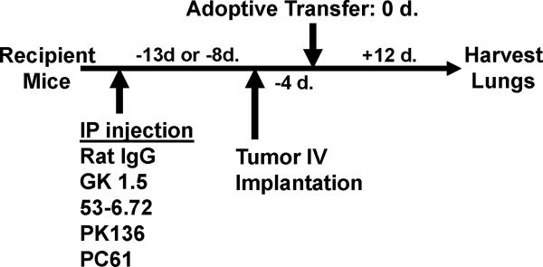
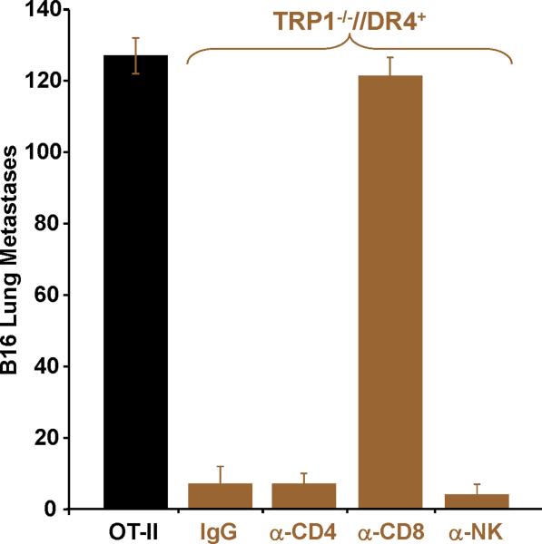
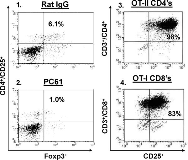
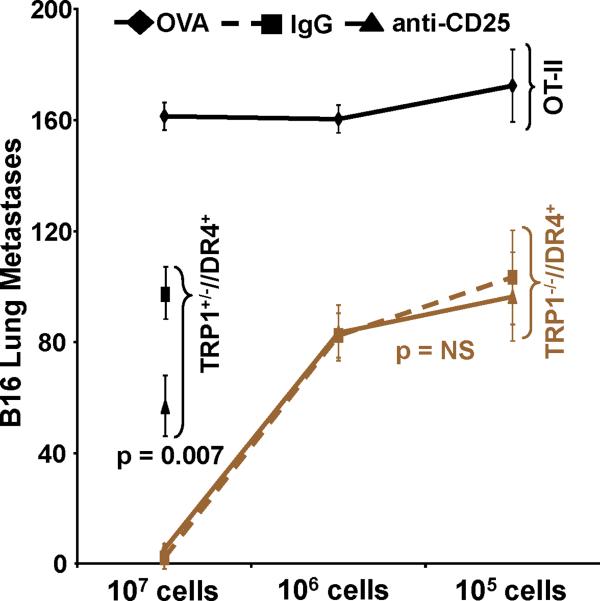
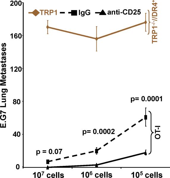
Despite progress made over the past 25 years, existing immunotherapies have limited clinical effectiveness in patients with cancer. Immune tolerance consistently blunts the generated immune response, and the largely solitary focus on CD8+ T cell immunity has proven ineffective in the absence of CD4+ T cell help. To address these twin-tier deficiencies, we developed a translational model of melanoma immunotherapy focused on the exploitation of high-avidity CD4+ T cells that become generated in germline antigen-deficient mice. We had previously identified a tyrosinase-related protein-1 specific HLA-DRB1*0401-restricted epitope. Using this epitope in conjunction with a newly described tyrosinase-related protein-1 germline-knockout, we demonstrate that endogenous tyrosinase-related protein-1 expression alters the functionality of the autoreactive T cell repertoire. More importantly, we show, by using major histocompatibility complex-mismatched combinations, that CD4+ T cells derived from the self-antigen deficient host indirectly triggers the eradication of established B16 lung metastases. We demonstrate that the treatment effect is mediated entirely by endogenous CD8+ T cells and is not affected by the depletion of host regulatory T cells. These findings suggest that high-avidity CD4+ T cells can overcome endogenous conditions and mediate their antitumor effects exclusively through the elicitation of CD8+ T cell immunity.


Figures

Figure 1
Figure 1
Despite differences between human and murine TRP-1, TRP-1277-297 is a potent self-antigen. (A) Sequence comparison analysis between human and murine TRP-1277-297 demonstrate conserved differences in positions 291 and 295, that involve aspartic acid (D) to glutamic acid (E) changes in both positions. (B) The IC50 nM concentration of the human TRP-1 variant is nearly one-third that of the murine form, but greater than epitopes that HA306-318 and gp10044-59. (C) Human TRP-1277-297 is a potent auto-antigen. DR4 Tg mice were immunized twice with mTRP-1 protein and extracted LN cells were stimulated with hTRP-1277-297 and expanded with IL-2. 14 days after restimulation cells were assayed for specific IFN-γ secretion by ELISA. Immune responses were superior to hTRP-1 peptide (pulsed onto DR4+ 1088 EBV-B at 50 μM) and hTRP-1 protein (pulsed onto DR4+ 1088 EBV-B at 25 μg/ml) when compared with reactivities observed to mTRP-1 peptide and protein. No significant immune response was observed to the control peptide HA306-318 or control protein OVA. All experiments were performed 2-3 times with similar results.
Figure 1
Figure 1
Despite differences between human and murine TRP-1, TRP-1277-297 is a potent self-antigen. (A) Sequence comparison analysis between human and murine TRP-1277-297 demonstrate conserved differences in positions 291 and 295, that involve aspartic acid (D) to glutamic acid (E) changes in both positions. (B) The IC50 nM concentration of the human TRP-1 variant is nearly one-third that of the murine form, but greater than epitopes that HA306-318 and gp10044-59. (C) Human TRP-1277-297 is a potent auto-antigen. DR4 Tg mice were immunized twice with mTRP-1 protein and extracted LN cells were stimulated with hTRP-1277-297 and expanded with IL-2. 14 days after restimulation cells were assayed for specific IFN-γ secretion by ELISA. Immune responses were superior to hTRP-1 peptide (pulsed onto DR4+ 1088 EBV-B at 50 μM) and hTRP-1 protein (pulsed onto DR4+ 1088 EBV-B at 25 μg/ml) when compared with reactivities observed to mTRP-1 peptide and protein. No significant immune response was observed to the control peptide HA306-318 or control protein OVA. All experiments were performed 2-3 times with similar results.
Figure 2
Figure 2
T cells from TRP1-/-//DR4+ Tg mice react more strongly to TRP-1 than wild-type controls. (A) TRP-1B-w (TRP-1-/-) mice were bred with DR4 Tg mice to generate TRP1-/-//DR4+ and control TRP1+/-//DR4+ littermates. (B) Flow cytometry analysis: LN populations obtained after vaccination are phenotypically equivalent. TRP1-/-//DR4+ (KO) and TRP1+/-//DR4+ (WT) littermates were immunized twice with recombinant hTRP-1. 14 days after the second immunization, LN cells (WT, panels 1-3; KO, panels 4-6) were harvested and stained with antibodies specific for murine CD4, CD8, CD3, CD25, and Foxp3. The panels are representative of three distinct experiments. (C) LN cells obtained from TRP-1 KO mice are more potent than WT controls. Following immunization, LN cells (4×10⁵ per well) were stimulated ex vivo in ELISPOT plates with soluble anti-CD3 (2 μg/ml) or DR4+ EBV-B cells (1×10⁵ per well; 1088 EBV-B) pulsed with peptide (100 □M) or protein (50 μg/ml) or lysate (105 cell equivalents). LN cells from both groups produce IFN-γ to anti-CD3 and EBV-B cells pulsed with mTRP-1277-297 or hTRP-1277-297 peptide, hTRP-1 or mTRP-1 protein, and to B16 and SK23 Mel lysate. Specific reactivity from both groups was blocked with anti-DR antibody HB55, but not with anti-MHC class I antibody W6/32. No reactivity observed to either the control peptide (HA306-318), protein (OVA) or lysates (MC-38, or 1102 Mel, both negative for TRP-1). (D) Ex Vivo ELISPOT titration assay: hTRP-1277-297-specific T cells from TRP1-/-//DR4+ mice are present at a higher frequency. TRP1-/-//DR4+ and TRP1+/-//DR4+ Tg littermates were immunized with hTRP-1 protein using the same immunization regimen. LN cells were harvested and stimulated (4×10⁵ per well in triplicates) ex vivo with hTRP-1277-297 pulsed onto DR4+ EBV-B cells (1×10⁵ per well; 1088 EBV-B) at titrating (100 to 0.0001 mM) peptide concentrations. All experiments were performed 2-3 times with similar results.
Figure 2
Figure 2
T cells from TRP1-/-//DR4+ Tg mice react more strongly to TRP-1 than wild-type controls. (A) TRP-1B-w (TRP-1-/-) mice were bred with DR4 Tg mice to generate TRP1-/-//DR4+ and control TRP1+/-//DR4+ littermates. (B) Flow cytometry analysis: LN populations obtained after vaccination are phenotypically equivalent. TRP1-/-//DR4+ (KO) and TRP1+/-//DR4+ (WT) littermates were immunized twice with recombinant hTRP-1. 14 days after the second immunization, LN cells (WT, panels 1-3; KO, panels 4-6) were harvested and stained with antibodies specific for murine CD4, CD8, CD3, CD25, and Foxp3. The panels are representative of three distinct experiments. (C) LN cells obtained from TRP-1 KO mice are more potent than WT controls. Following immunization, LN cells (4×10⁵ per well) were stimulated ex vivo in ELISPOT plates with soluble anti-CD3 (2 μg/ml) or DR4+ EBV-B cells (1×10⁵ per well; 1088 EBV-B) pulsed with peptide (100 □M) or protein (50 μg/ml) or lysate (105 cell equivalents). LN cells from both groups produce IFN-γ to anti-CD3 and EBV-B cells pulsed with mTRP-1277-297 or hTRP-1277-297 peptide, hTRP-1 or mTRP-1 protein, and to B16 and SK23 Mel lysate. Specific reactivity from both groups was blocked with anti-DR antibody HB55, but not with anti-MHC class I antibody W6/32. No reactivity observed to either the control peptide (HA306-318), protein (OVA) or lysates (MC-38, or 1102 Mel, both negative for TRP-1). (D) Ex Vivo ELISPOT titration assay: hTRP-1277-297-specific T cells from TRP1-/-//DR4+ mice are present at a higher frequency. TRP1-/-//DR4+ and TRP1+/-//DR4+ Tg littermates were immunized with hTRP-1 protein using the same immunization regimen. LN cells were harvested and stimulated (4×10⁵ per well in triplicates) ex vivo with hTRP-1277-297 pulsed onto DR4+ EBV-B cells (1×10⁵ per well; 1088 EBV-B) at titrating (100 to 0.0001 mM) peptide concentrations. All experiments were performed 2-3 times with similar results.
Figure 3
Figure 3
CD4+ T cells derived from TRP-1 KO mice recognize tumor in vitro. TRP1-/-//DR4+and TRP1+/-//DR4+ Tg littermates were immunized with hTRP-1 protein and expanded in vitro. (A) Flow cytometric analysis of experimental T cell populations. Both groups of in vitro IL-2 expanded WT and KO T cells are equivalently CD4high/CD8dim (panels 1 and 3) and Treg negative CD4high/CD25high/Foxp3dim (panel 2 and 4). The panels are representative of three distinct experiments. (B) Significant differences in in vitro reactivity to titering concentrations of hTRP-1277-297 pulsed onto DR4+ EBV-B cells (1088 EBV-B) were observed between groups. (C) CD4+ T cells derived from germline-deficient hosts strongly recognize intact melanoma. CD4+ T cells from both groups differentially react to both the TRP-1 peptide and the recombinant protein when pulsed on DR4+ targets (DR4 Tg splenocytes), but not when pulsed onto DR4- APCs (C57BL/6 splenocytes, I-Ab+). No reactivity was observed to control protein OVA. Specific reactivity was also observed to both HLA-DR4+ and TRP-1+ human melanomas (526 and 624 Mel pretreated with IFN-γ and 1088 Mel stably transduced with CIITA) and to murine melanoma B16-DR4 (B16 stably transduced with HLA-DRB1*0401). No reactivity was observed against control tumor B16 (DR4-). Recognition of B16-DR4 was blocked with mAb HB55 (anti-class II), but not with W6/32 (anti-class I). (D) CD4+ T cells from both groups exhibit a Th1 cytokine profile (IFN-γ production, but none to IL-4 or IL-17A by ELISA) in response to specific peptide and tumor (B16-DR4, but not MC-38-DR4). All experiments were performed 2-3 times with similar results.
Figure 3
Figure 3
CD4+ T cells derived from TRP-1 KO mice recognize tumor in vitro. TRP1-/-//DR4+and TRP1+/-//DR4+ Tg littermates were immunized with hTRP-1 protein and expanded in vitro. (A) Flow cytometric analysis of experimental T cell populations. Both groups of in vitro IL-2 expanded WT and KO T cells are equivalently CD4high/CD8dim (panels 1 and 3) and Treg negative CD4high/CD25high/Foxp3dim (panel 2 and 4). The panels are representative of three distinct experiments. (B) Significant differences in in vitro reactivity to titering concentrations of hTRP-1277-297 pulsed onto DR4+ EBV-B cells (1088 EBV-B) were observed between groups. (C) CD4+ T cells derived from germline-deficient hosts strongly recognize intact melanoma. CD4+ T cells from both groups differentially react to both the TRP-1 peptide and the recombinant protein when pulsed on DR4+ targets (DR4 Tg splenocytes), but not when pulsed onto DR4- APCs (C57BL/6 splenocytes, I-Ab+). No reactivity was observed to control protein OVA. Specific reactivity was also observed to both HLA-DR4+ and TRP-1+ human melanomas (526 and 624 Mel pretreated with IFN-γ and 1088 Mel stably transduced with CIITA) and to murine melanoma B16-DR4 (B16 stably transduced with HLA-DRB1*0401). No reactivity was observed against control tumor B16 (DR4-). Recognition of B16-DR4 was blocked with mAb HB55 (anti-class II), but not with W6/32 (anti-class I). (D) CD4+ T cells from both groups exhibit a Th1 cytokine profile (IFN-γ production, but none to IL-4 or IL-17A by ELISA) in response to specific peptide and tumor (B16-DR4, but not MC-38-DR4). All experiments were performed 2-3 times with similar results.
Figure 4
Figure 4
Adoptively transferred CD4+ T cells derived from TRP-1 KO mice eradicate established tumor by priming host CD8+ T cells. To prepare cells for adoptive transfer, TRP1-/-//DR4+ and TRP1+/-//DR4+ Tg littermates were immunized with hTRP-1 protein and expanded in vitro. Control OT-II mice were similarly immunized and expanded. (A) A general schematic of all tumor treatment experiments. (B) Treatment is antigen-specific and DR4-restricted. Expanded T cells were adoptively transferred (107 per mouse) into three different tumor-bearing hosts at 5 mice per group: 1) B16 (TRP-1+) lung metastases in DR4 transgenics (DR4+); 2) B16 lung metastases in C57BL/6 mice (DR4-); 3) MC38 (TRP-1-) lung metastases in DR4 transgenics. Each group had been previously IV tail vein inoculated with tumor. CD4+ T cells derived from the TRP-1-/-//DR4+ mice nearly fully eradicate the 4-day lung metastases (mean = 5; p = 0.0006 using a two-tailed Student T-test) when compared to littermate (WT) controls (mean = 68) and control OVA-specific CD4+ T cells (mean: 156). No treatment effect was observed (mean = >150 lung metastases for all treatment groups) in mice bearing the control tumor cell line (MC38) or in recipient mice lacking the specific restriction element (C57BL/6 mice; DR4-). (C) Treatment of B16 lung metastases is dependent upon host CD8+ T cells. Tumor bearing (B16 only) DR4 Tg mice were depleted of individual effector-cell subsets (CD4, CD8, NK and NKT) following IP injection of mAb's prior to adoptive transfer of CD4+ T cells derived from TRP1-/-//DR4+ mice. Control OVA-specific CD4+ T cells were used to measure the non-specific effects of adoptive transfer. Each experimental arm involved 5 mice per group and all experiments were performed 2-3 times with similar results.
Figure 4
Figure 4
Adoptively transferred CD4+ T cells derived from TRP-1 KO mice eradicate established tumor by priming host CD8+ T cells. To prepare cells for adoptive transfer, TRP1-/-//DR4+ and TRP1+/-//DR4+ Tg littermates were immunized with hTRP-1 protein and expanded in vitro. Control OT-II mice were similarly immunized and expanded. (A) A general schematic of all tumor treatment experiments. (B) Treatment is antigen-specific and DR4-restricted. Expanded T cells were adoptively transferred (107 per mouse) into three different tumor-bearing hosts at 5 mice per group: 1) B16 (TRP-1+) lung metastases in DR4 transgenics (DR4+); 2) B16 lung metastases in C57BL/6 mice (DR4-); 3) MC38 (TRP-1-) lung metastases in DR4 transgenics. Each group had been previously IV tail vein inoculated with tumor. CD4+ T cells derived from the TRP-1-/-//DR4+ mice nearly fully eradicate the 4-day lung metastases (mean = 5; p = 0.0006 using a two-tailed Student T-test) when compared to littermate (WT) controls (mean = 68) and control OVA-specific CD4+ T cells (mean: 156). No treatment effect was observed (mean = >150 lung metastases for all treatment groups) in mice bearing the control tumor cell line (MC38) or in recipient mice lacking the specific restriction element (C57BL/6 mice; DR4-). (C) Treatment of B16 lung metastases is dependent upon host CD8+ T cells. Tumor bearing (B16 only) DR4 Tg mice were depleted of individual effector-cell subsets (CD4, CD8, NK and NKT) following IP injection of mAb's prior to adoptive transfer of CD4+ T cells derived from TRP1-/-//DR4+ mice. Control OVA-specific CD4+ T cells were used to measure the non-specific effects of adoptive transfer. Each experimental arm involved 5 mice per group and all experiments were performed 2-3 times with similar results.
Figure 5
Figure 5
Adoptively transferred CD4+ T cells derived from TRP-1 KO's are maximally effective against tumor regardless of Treg status. (A) Flow cytometric analysis obtained from peripheral blood samples on the day of adoptive transfer of host Treg status (CD4+/CD25+/Foxp3+) following depletion with control rat IgG (panel 1) or with PC61 (panel 2), respectively. Flow cytometric analysis of control in vitro expanded OT-II and OT-I cells are CD4high/CD25high (panel 3) and CD8high/CD25high (panel 4), respectively; the panels are representative of three distinct experiments. (B) Treatment of B16 lung metastases is unaffected by Treg depletion. Tumor bearing (B16 only) DR4 Tg mice were depleted of Tregs following IP injection of mAb PC61 prior to adoptive transfer. CD4+ T cells derived from KO mice and control OT-II mice were transferred at titering concentrations (107, 106, and 105). CD4+ T cells derived from TRP-1 WT mice were transferred at 107 cells only. Pre-treatment with PC61 has no impact on treatment when compared with control rat IgG administration using KO cells when compared with WT cells. No significant treatment effect was observed using the control OVA-specific CD4+ T cell transfer. (C) OVA-specific CD8+/CD25+ T cells more effectively eradicate established tumor in hosts depleted of Tregs. Tumor bearing (E.G7 only) C57BL/6 mice were similarly depleted of Tregs prior to adoptive transfer. CD8+ T cells derived from OT-I mice and control TRP-1-specific CD4's were adoptively transferred at titering concentrations. Pre-treatment with PC61 significantly improved the overall treatment effect at both 106 and 105 cells infused when compared with control rat IgG. No significant treatment effect was observed using control TRP-1-specific CD4+ T cells. Each experimental arm involved 5 mice per group, and all experiments were performed at least 2-3 times with similar results.
Figure 5
Figure 5
Adoptively transferred CD4+ T cells derived from TRP-1 KO's are maximally effective against tumor regardless of Treg status. (A) Flow cytometric analysis obtained from peripheral blood samples on the day of adoptive transfer of host Treg status (CD4+/CD25+/Foxp3+) following depletion with control rat IgG (panel 1) or with PC61 (panel 2), respectively. Flow cytometric analysis of control in vitro expanded OT-II and OT-I cells are CD4high/CD25high (panel 3) and CD8high/CD25high (panel 4), respectively; the panels are representative of three distinct experiments. (B) Treatment of B16 lung metastases is unaffected by Treg depletion. Tumor bearing (B16 only) DR4 Tg mice were depleted of Tregs following IP injection of mAb PC61 prior to adoptive transfer. CD4+ T cells derived from KO mice and control OT-II mice were transferred at titering concentrations (107, 106, and 105). CD4+ T cells derived from TRP-1 WT mice were transferred at 107 cells only. Pre-treatment with PC61 has no impact on treatment when compared with control rat IgG administration using KO cells when compared with WT cells. No significant treatment effect was observed using the control OVA-specific CD4+ T cell transfer. (C) OVA-specific CD8+/CD25+ T cells more effectively eradicate established tumor in hosts depleted of Tregs. Tumor bearing (E.G7 only) C57BL/6 mice were similarly depleted of Tregs prior to adoptive transfer. CD8+ T cells derived from OT-I mice and control TRP-1-specific CD4's were adoptively transferred at titering concentrations. Pre-treatment with PC61 significantly improved the overall treatment effect at both 106 and 105 cells infused when compared with control rat IgG. No significant treatment effect was observed using control TRP-1-specific CD4+ T cells. Each experimental arm involved 5 mice per group, and all experiments were performed at least 2-3 times with similar results.

References

    1. Boon T, Coulie PG, Van Den Eynde BJ, van der BP. Human T cell responses against melanoma. Annu.Rev.Immunol. 2006;24:175–208. - PubMed
    1. Rosenberg SA, Restifo NP, Yang JC, Morgan RA, Dudley ME. Adoptive cell transfer: a clinical path to effective cancer immunotherapy. Nat.Rev.Cancer. 2008;8:299–308. - PMC - PubMed
    1. Peggs KS, Segal NH, Allison JP. Targeting immunosupportive cancer therapies: accentuate the positive, eliminate the negative. Cancer Cell. 2007;12:192–199. - PubMed
    1. Rosenberg SA, Yang JC, Restifo NP. Cancer immunotherapy: moving beyond current vaccines. Nat.Med. 2004;10:909–915. - PMC - PubMed
    1. Lizee G, Cantu MA, Hwu P. Less yin, more yang: confronting the barriers to cancer immunotherapy. Clin.Cancer Res. 2007;13:5250–5255. - PubMed

Publication types

MeSH terms