. 2009 Jul 30;460(7255):642-6.
doi: 10.1038/nature08139. Epub 2009 Jun 28.

MicroRNA-mediated switching of chromatin-remodelling complexes in neural development

Affiliations

MicroRNA-mediated switching of chromatin-remodelling complexes in neural development

Andrew S Yoo et al. Nature. .

Erratum in

  • Nature. 2009 Sep 10;461(7261):296

Abstract

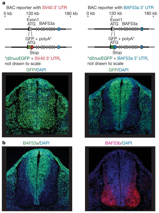
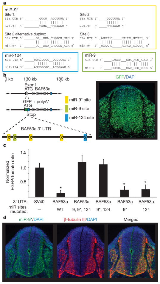
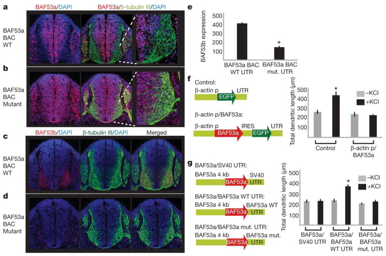
One of the most distinctive steps in the development of the vertebrate nervous system occurs at mitotic exit when cells lose multipotency and begin to develop stable connections that will persist for a lifetime. This transition is accompanied by a switch in ATP-dependent chromatin-remodelling mechanisms that appears to coincide with the final mitotic division of neurons. This switch involves the exchange of the BAF53a (also known as ACTL6a) and BAF45a (PHF10) subunits within Swi/Snf-like neural-progenitor-specific BAF (npBAF) complexes for the homologous BAF53b (ACTL6b) and BAF45b (DPF1) subunits within neuron-specific BAF (nBAF) complexes in post-mitotic neurons. The subunits of the npBAF complex are essential for neural-progenitor proliferation, and mice with reduced dosage for the genes encoding its subunits have defects in neural-tube closure similar to those in human spina bifida, one of the most serious congenital birth defects. In contrast, BAF53b and the nBAF complex are essential for an evolutionarily conserved program of post-mitotic neural development and dendritic morphogenesis. Here we show that this essential transition is mediated by repression of BAF53a by miR-9* and miR-124. We find that BAF53a repression is mediated by sequences in the 3' untranslated region corresponding to the recognition sites for miR-9* and miR-124, which are selectively expressed in post-mitotic neurons. Mutation of these sites led to persistent expression of BAF53a and defective activity-dependent dendritic outgrowth in neurons. In addition, overexpression of miR-9* and miR-124 in neural progenitors caused reduced proliferation. Previous studies have indicated that miR-9* and miR-124 are repressed by the repressor-element-1-silencing transcription factor (REST, also known as NRSF). Indeed, expression of REST in post-mitotic neurons led to derepression of BAF53a, indicating that REST-mediated repression of microRNAs directs the essential switch of chromatin regulatory complexes.


Figures

Figure 1
Figure 1. BAF53a repression is mediated by sequences within its 3′ UTR
a, Schematic diagrams of BAC clones of approximately 180 kb containing BAF53a and the BAC-based reporter constructs. Exon 1 from the ATG of BAF53a to the end of the exon was replaced by d2nucEGFP and 3′ UTR. Whereas EGFP failed to be repressed in neurons with SV40 UTR (lower-left panel), the BAF53a 3′ UTR allowed efficient downregulation of the reporter in neurons (lower-right panel). Photographs in the lower panels show transverse sections of the neural tubes of E11.5 transgenic embryos. DAPI, 4,6-diamidino-2-phenylindole. b, BAF53a and BAF53b are expressed selectively in progenitors (green) and neurons (red), respectively, in the neural tubes of E11.5 embryos.
Figure 2
Figure 2. BAF53a is a target of miR-9* and miR-124
a, Predicted configurations of duplex formations between miRNAs and their target sites. b, Expression of BAF53a BAC reporter, containing mutations at the miRNA-binding sites in the 3′ UTR of d2nucEGFP. Point mutations were made to 4–6 bp predicted to bind to the 5′ ends of miR-9, miR-9* and miR-124. The photograph shows a representative expression pattern seen in three individual transgenic lines. c, BAF53a 3′ UTR sensor experiments in stable Chinese hamster ovary (CHO) cells expressing miR-9, mirR-9* and miR-124 and sensor reporter constructs. The quantification of sensor expression was expressed as the ratio of EGFP/Tomato signals and normalized to the ratio values of the SV40 UTR sensor. Error bars, s.d. Signal intensities were averaged in ten random fields totalling approximately 500–1,000 cells. *P <0.001, Student’s t-test. WT, wild type. d, Selective expression of miR-9* in post-mitotic neurons. Photographs show fluorescent in situ hybridization analysis of miR-9* expression (in green) and β-tubulin-III staining (in red) in the neural tubes of E11.5 embryos.
Figure 3
Figure 3. BAF53a repression is essential for activity-dependent dendritic outgrowth in neurons
a, Normal downregulation of BAF53a in post-mitotic neurons in transgenic embryos with wild type BAF53a BAC. The rightmost panel shows the lower-right quadrant of the neural tube. b, Persistent expression of BAF53a in neurons seen with BAF53a BAC containing point mutations in the miRNA-binding sites. c, Normal expression of BAF53b (red) in β-tubulin-III-positive (green) neurons in transgenic embryos with wild-type BAF53a BAC. d, Reduced BAF53b expression with persistent expression of BAF53a in neurons. e, Quantification of BAF53b expression: ratio of BAF53b level (arbitrary units) and β-tubulin-III-positive neurons. Average values are from eight sections of the neural tube. Error bars, s.e. *P <0.005, Student’s t-test. f, Constructs to overexpress BAF53a in cultured hippocampal neurons and quantification of dendritic outgrowth of GFP-positive neurons upon stimulation using KCl. The average values are from five individual coverslips from two independent experiments, with each coverslip containing 50–100 scored neurons. Error bars, s.e. *P <0.005, Student’s t-test. p, promoter; IRES, internal ribosome entry site. g, Schematic diagrams of BAF53a expression constructs using different 3′ UTRs and quantification of dendritic outgrowth of transfected neurons upon stimulation using KCl. In independent experiments, we found that the 4-kb upstream region of BAF53a (illustrated) was sufficient to drive expression of GFP reporters that could be repressed by endogenous miR-9* and miR-124. Error bars, s.e. *P <0.001, Student’s t-test.
Figure 4
Figure 4. Effect of miR-9* and miR-124 overexpression in progenitors and REST in neurons
a, miR-9* and miR-124 overexpression constructs (miR-9*-124) expressed in progenitors. nucEGFP, nuclear enhanced green fluorescent protein. b, Proliferative defect caused by miR-9* and miR-124 overexpression in progenitors. Overexpression of miR-9* and miR-124 reduced BAF53a expression in the EGFP-positive progenitors relative to control (upper-right graph). Photographs show transgenic embryos containing either control or miR-9*-124 overexpression constructs and Ki-67 staining, quantified in the lower-right graph on the basis of nine individual tissue sections. Error bars, s.e. *P <0.001, Student’s t-test. c, BAF53a regulation is mediated by REST. Hippocampal neurons cultured from E18.5 embryos were transfected with control (EGFP only), REST (EGFP +REST expression constructs) or REST +miR-9*-124 (EGFP +REST +miRNA overexpression constructs). The quantification represents average percentages of transfected GFP-positive neurons that had BAF53a expression from five coverslips. We analysed 15–25 GFP-positive neurons per coverslip. Error bars, s.e. *P <0.05, Student’s t-test. d, Genetic pathway leading to BAF53-subunit switching during neuronal differentiation.

References

    1. Noctor SC, Martinez-Cerdeno V, Ivic L, Kriegstein AR. Cortical neurons arise in symmetric and asymmetric division zones and migrate through specific phases. Nature Neurosci. 2004;7:136–144. - PubMed
    1. Temple S. The development of neural stem cells. Nature. 2001;414:112–117. - PubMed
    1. Lessard J, et al. An essential switch in subunit composition of a chromatin remodeling complex during neural development. Neuron. 2007;55:201–215. - PMC - PubMed
    1. Wu J, et al. Regulation of dendritic development by neuron-specific chromatin remodeling complexes. Neuron. 2007;56:94–108. - PubMed
    1. Parrish JZ, Kim MD, Jan LY, Jan YN. Genome-wide analyses identify transcription factors required for proper morphogenesis of Drosophila sensory neuron dendrites. Genes Dev. 2006;20:820–835. - PMC - PubMed

Publication types

MeSH terms