. 2009 Aug;12(8):988-95.
doi: 10.1038/nn.2358. Epub 2009 Jun 28.

A discrete alcohol pocket involved in GIRK channel activation

Affiliations

A discrete alcohol pocket involved in GIRK channel activation

Prafulla Aryal et al. Nat Neurosci. 2009 Aug.

Abstract

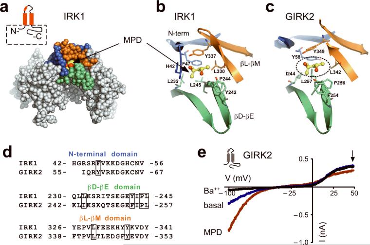
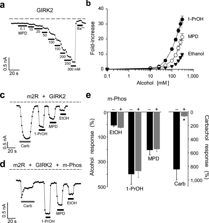
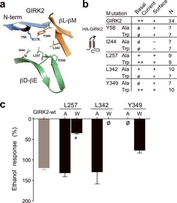
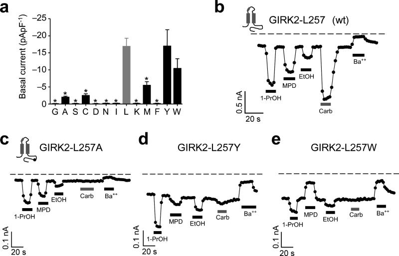
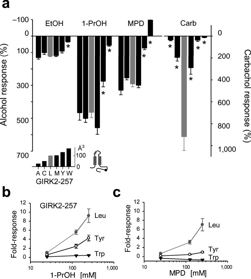
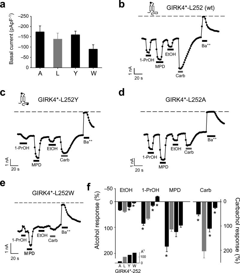
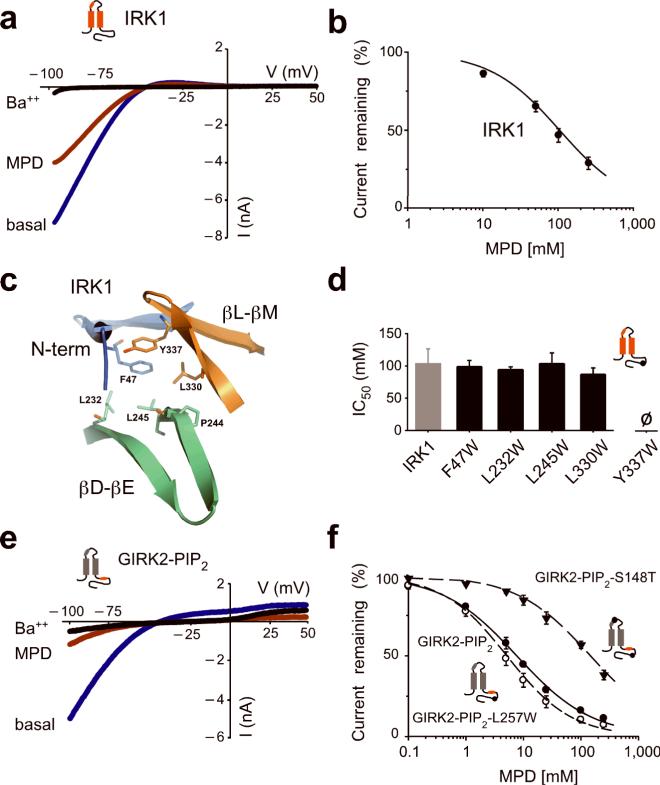
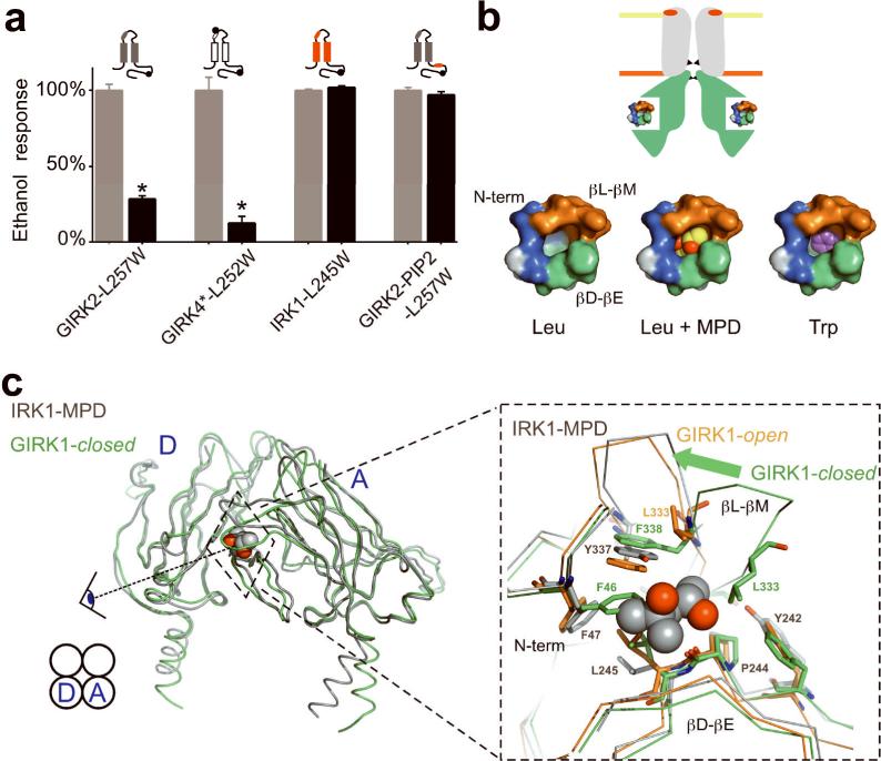
Ethanol modifies neural activity in the brain by modulating ion channels. Ethanol activates G protein-gated inwardly rectifying K(+) channels, but the molecular mechanism is not well understood. Here, we used a crystal structure of a mouse inward rectifier containing a bound alcohol and structure-based mutagenesis to probe a putative alcohol-binding pocket located in the cytoplasmic domains of GIRK channels. Substitutions with bulkier side-chains in the alcohol-binding pocket reduced or eliminated activation by alcohols. By contrast, alcohols inhibited constitutively open channels, such as IRK1 or GIRK2 engineered to strongly bind PIP(2). Mutations in the hydrophobic alcohol-binding pocket of these channels had no effect on alcohol-dependent inhibition, suggesting an alternate site is involved in inhibition. Comparison of high-resolution structures of inwardly rectifying K(+) channels suggests a model for activation of GIRK channels using this hydrophobic alcohol-binding pocket. These results provide a tool for developing therapeutic compounds that could mitigate the effects of alcohol.


Figures

Fig. 1
Fig. 1. A conserved alcohol-binding pocket in IRK1 and GIRK2 channels
a) CPK representation of the cytoplasmic domains from two subunits of IRK1 in complex with an alcohol, MPD (PDB: 2GIX). The pocket for MPD is formed by three structural elements: the N-terminal domain (blue) and the βL-βM ribbon (orange) from one subunit, and the βD-βE ribbon (green) from an adjacent subunit. Inset, schematic of IRK1 (red) shows the major structural elements of the subunit including pore loop and helix, two transmembrane domains, and N- and C-terminals used in the structure (dashed box). b,c) Detailed structural views of amino acids forming the hydrophobic alcohol pocket of IRK1 with MPD (ball and stick) (b) and a putative hydrophobic alcohol pocket in GIRK2 (PDB: 2E4F) (c). Amino acid residues shown in stick format are colored according to the domain they originate from; as MPD is shown in ball-and-stick format. The putative position of MPD in the GIRK2 (dashed circle) was obtained by superposition of two adjacent cytoplasmic domains from IRK1 structure and corresponding subunits from GIRK2 structure. d) Sequence alignment for the three domains comprising the hydrophobic alcohol pocket in IRK1 and GIRK2 channels. Boxes indicate amino acids that form hydrophobic and hydrogen-bond interactions in IRK1-MPD, and are conserved in GIRK2. ‘HG’ in the N-terminal domain of IRK1 originates from the polypeptide linker in the IRK1-MPD structure. e) Current-voltage plots for GIRK2 channels recorded in the presence of 20K (blue), 20K plus 1 mM Ba++ (black) or 20K plus 100 mM MPD (red). Currents were elicited by voltage ramps from −100 mV to +50 mV. MPD-induced current was 246% ± 27% (n=5, mean and s.e.m.) of basal K+ current (Ba++ sensitive).
Fig. 2
Fig. 2. MPD activates GIRK2 in a manner similar to other alcohols
a) The inward current through GIRK2 channels plotted as a function of time (at −100 mV) shows the response to the increasing concentrations of MPD and to 1 mM Ba++. Dashed line shows zero current level. b) Dose-response curves are shown for MPD (n=6), 1-PrOH (n=6), and EtOH (n=6). The fold-increase was calculated by normalizing to the basal K+ current (Ba++-sensitive). c-e) Chelating Gβγ with m-Phos attenuates m2R- but not alcohol-mediated activation of GIRK2. Current responses recorded at −100 mV are shown for m2R/GIRK2 (c) or m2R/GIRK2/m-Phos (d) in response to 100 mM 1-PrOH, 100 mM MPD, 100 mM EtOH, or 5 μM carbachol. e) Bar graphs show the mean percentage alcohol and carbachol responses (± s.e.m.), normalized to the Ba++-sensitive basal current, in the absence (solid, n=4) or presence of m-Phos (grey, n=7). Asterisks indicate statistical significant difference from wild-type (P < 0.05).
Fig. 3
Fig. 3. Ala/Trp scan of the hydrophobic alcohol-binding pocket in GIRK2
a) Ribbon structure shows amino acids that line the hydrophobic alcohol pocket in GIRK2. b) Summary table of Ala/Trp mutagenesis. Basal K+ currents (Ba++-sensitive) were divided into three groups; < −1 pApF−1 (ø), −1 to −5 pApF−1 (+) and > −5 pApF−1 (++) (n = number of recordings). Surface expression on the plasma membrane was assessed in separate experiments with HA-tagged channels; detected on the surface (+) or detected only in cytoplasm (−). See Supplemental Fig. S1. Schematic shows location of HA tag (‘v’) in GIRK2 (grey). c) Bar graph shows the mean ethanol percentage response, normalized to the basal K+ current, for different mutant channels (± s.e.m.). L257W showed a significant statistical decrease in EtOH response (*P < 0.05 vs. wild-type).
Fig. 4
Fig. 4. Comprehensive mutagenesis of GIRK2-L257 in βD-βE ribbon of hydrophobic alcohol-binding pocket reveals changes in alcohol- and Gβγ-activated currents
a) Bar graph shows the mean (± s.e.m.) amplitude of basal K+ current (Ba++-sensitive) for substitutions of increasing molecular side-chain volume at GIRK2-L257: Gly (n=7), Ala (n=9), Ser (n=7), Cys (n=8), Asp (n=7), Asn (n=6), Ile (n=7), Leu (wt; n=34; grey bar), Lys (n=7), Met (n=8), Phe (n=7), Tyr (n=9) and Trp (n=9). b-e) Inward K+ currents for wild-type GIRK2 (b) and the indicated GIRK2-L257 mutants (c-e) in response to 100 mM 1-PrOH, 100 mM MPD, 100 mM EtOH, 5 μM carbachol, or 1 mM Ba++. Inset shows the approximate position of the C-terminal mutation.
Fig. 5
Fig. 5. Reduced alcohol activation with increasing bulkiness of amino acid substitutions at GIRK2-L257
a) Bar graph shows the mean percentage response to different alcohols and carbachol (± s.e.m.), normalized to the basal K+ current (Ba++-sensitive). Upward response indicates inhibition. Amino acid substitutions are arranged by increasing side-chain volume (Å3, see inset). Asterisk indicates significant statistical difference (P < 0.05 vs. Leu). b,c) Dose-response curves are shown for GIRK2-L257, GIRK2-L257Y and GIRK2-L257W channels for 1-PrOH (b) and MPD (c). Note suppression of alcohol activation at all concentrations tested.
Fig. 6
Fig. 6. Mutations in the hydrophobic alcohol-binding pocket of GIRK4* alter alcohol-activated currents
a) Mean basal K+ currents (Ba++-sensitive) measured for Ala (n=8), Leu (wt; grey bar, n=8), Tyr (n=8), and Trp (n=8) substitutions at GIRK4*-L252. There are no statistical differences in basal currents (P > 0.05 vs. Leu). b-e) Inward K+ currents for GIRK4* (b) and different GIRK4*-L252 mutants (c-e) in response to 100 mM 1-PrOH, 100 mM MPD, 100 mM EtOH, 5 μM carbachol, or 1 mM Ba++. f) Bar graphs show the mean percentage responses to different alcohols and carbachol, normalized to the basal K+ current (Ba++-sensitive). Amino acid substitutions are arranged by increasing side-chain volume (Å3) (see inset). Asterisk indicates significant statistical difference (P < 0.05 vs. Leu). Channel schematics show the approximate position of the pore-helix (white ellipse) mutation, for making GIRK4*, and the C-terminal mutation (black circle). All values are mean ± s.e.m.
Fig. 7
Fig. 7. Mutations in the hydrophobic alcohol-binding pocket of IRK1 have no effect on alcohol-dependent inhibition
a) Current-voltage plots for IRK1 channels are shown for 20K (blue), 20K plus 1 mM Ba++ (black) or 20K plus 100 mM MPD (red). MPD inhibited the basal K+ current (Ba++-sensitive) by 53.1%± 4.1% (n=8). b) Dose-response curve is shown for MPD inhibition of IRK1 channel. Smooth curve shows best fit using the Hill equation, with an IC50 of 104 ± 23 mM and Hill coefficient of 0.93 ± 0.03 (n=8). c) Structural view of amino acids that line the hydrophobic alcohol pocket in IRK1. d) Bar graph shows mean IC50‘s for MPD-dependent inhibition of IRK1 (n=8), IRK1-F47W (n=7), IRK1-L232W (n=7), IRK1-L245W (n=6), IRK1-L330W (n=6). There is no statistical difference compared to wild-type IRK1 (P > 0.05). e) Current-voltage plots are shown for GIRK2-PIP2 (GIRK2 engineered with high affinity PIP2 binding domain from IRK1) channels recorded in the presence of 20K (blue), 20K plus 1 mM Ba++ (black) or 20K plus 100 mM MPD (red). f) Dose-response curves for MPD-dependent inhibition of GIRK2-PIP2 (solid circle), GIRK2-PIP2-L257W (open circle) and GIRK2-PIP2-S148T (solid triangle). Smooth curves show best fit using the Hill equation and having IC50‘s and Hill coefficients of 7.7 ± 1.0 mM and 0.66 ± 0.03 (n=5) for GIRK2-PIP2, 5.2 ± 1.0 mM and 0.77 ± 0.04 (n=5) for GIRK2-PIP2-L257W, and 147.0 ± 31.5 mM and 0.67 ± 0.05 (n=6) for GIRK2-PIP2–S148T. All values are mean ± s.e.m.
Fig. 8
Fig. 8. Model for alcohol-dependent activation of GIRK channels
a) Bar graph shows the mean percentage EtOH response (activation or inhibition normalized to wild-type) for a Trp mutation in four different channels, GIRK2-L257W (n=9), GIRK4-L252W (n=8), IRK1-L245W (n=8) and GIRK2-PIP2-L257W (n=5). Only mutations in alcohol-binding pocket of wild-type GIRK channels affect the response to alcohol. b) Top, schematic of inward rectifier shows location of alcohol-binding pocket in cytoplasmic domains, two gates (G-loop and M2 transmembrane; black triangles) and pore-helix region (red ellipse). PIP2 is enriched in lower leaflet of bilayer (orange). Below, molecular surface representations of the alcohol pocket without (Leu), with MPD (Leu+MPD) and modeled with L257W (Trp), using the IRK1-MPD structure as a guide. c) Left, alignment of the putative closed state of GIRK1 chimeric channel (GIRK1-closed; green) (PDB:2QKS) with the IRK1-MPD structure (grey) (PDB:2GIX). Spaghetti structures show two adjacent cytoplasmic subunits (subunits D and A) and the hydrophobic alcohol pocket at the cytoplasmic subunit interface. Right, zoom shows alignment of the N-terminal domain, βD-βE and βL-βM ribbons from the IRK1-MPD (grey), GIRK1-open (orange) and GIRK1-closed (green) structures. IRK1-MPD aligns better with the putative open state of GIRK1. Note the significant displacement in the βL-βM beta ribbon element (arrow) and the side-chains of hydrophobic amino acids in the two structures. GIRK1-closed but not GIRK1-open has a collapsed alcohol-binding pocket, due to interaction and rotation of F46 (IRK1-F47), L246 (IRK1-L245) and F338 (IRK1-Y337). GIRK1-L333 in the βL-βM domain, implicated previously in Gββ gating of GIRK channels-, is shown for reference.

Similar articles

Cited by

References

    1. Cardoso RA, Brozowski SJ, Chavez-Noriega LE, Harpold M, Valenzuela CF, Harris RA. Effects of ethanol on recombinant human neuronal nicotinic acetylcholine receptors expressed in Xenopus oocytes. J Pharmacol Exp Ther. 1999;289:774–780. - PubMed
    1. Lovinger DM, White G, Weight FF. Ethanol inhibits NMDA-activated ion current in hippocampal neurons. Science. 1989;243:1721–1724. - PubMed
    1. Mihic SJ, Ye Q, Wick MJ, Koltchine VV, Krasowski MD, Finn SE, Mascia MP, Valenzuela CF, Hanson KK, Greenblatt EP, Harris RA, Harrison NL. Sites of alcohol and volatile anaesthetic action on GABAA and glycine receptors. Nature. 1997;389:385–389. - PubMed
    1. Zhou Q, Lovinger DM. Pharmacologic characteristics of potentiation of 5-HT3 receptors by alcohols and diethyl ether in NCB-20 neuroblastoma cells. J Pharmacol Exp Ther. 1996;278:732–740. - PubMed
    1. Harris RA, Trudell JR, Mihic SJ. Ethanol's molecular targets. Sci Signal. 2008;1:re7. - PMC - PubMed

Publication types

MeSH terms

Substances