. 2009 Jul;16(7):698-703.
doi: 10.1038/nsmb.1607. Epub 2009 Jun 28.

Structural insights into hedgehog ligand sequestration by the human hedgehog-interacting protein HHIP

Affiliations

Structural insights into hedgehog ligand sequestration by the human hedgehog-interacting protein HHIP

Benjamin Bishop et al. Nat Struct Mol Biol. 2009 Jul.

Abstract

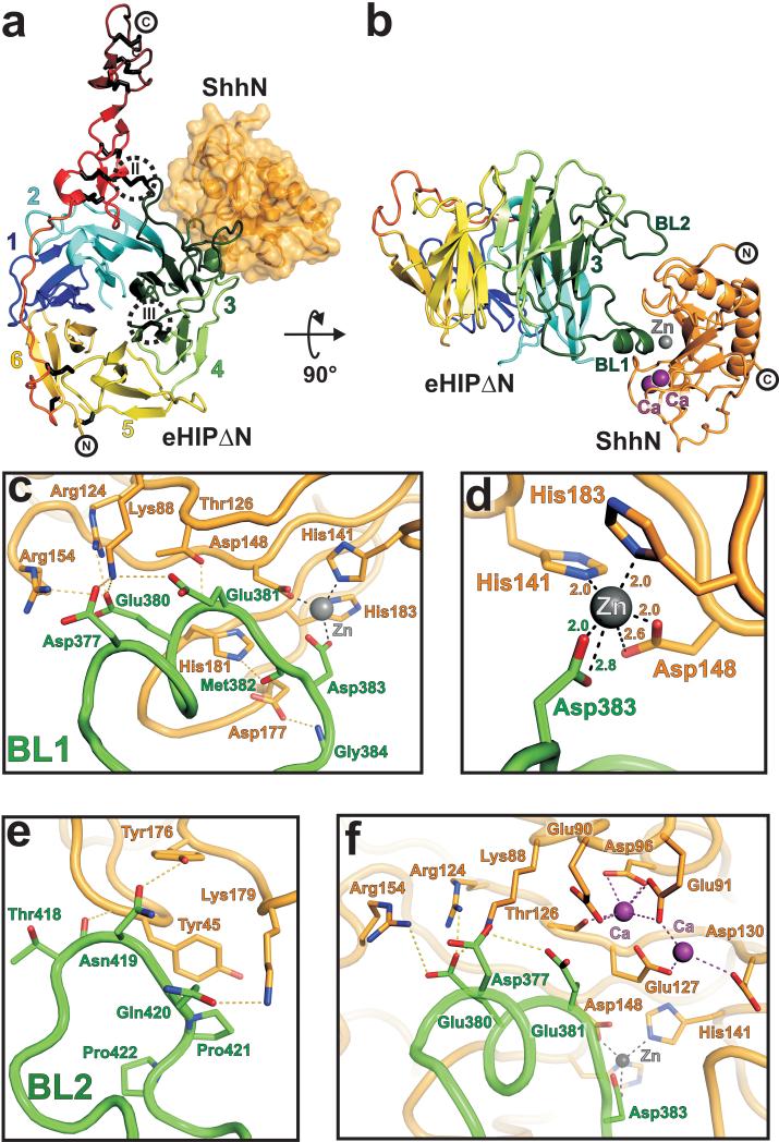
Hedgehog (Hh) morphogens have fundamental roles in development, whereas dysregulation of Hh signaling leads to disease. Multiple cell-surface receptors are responsible for transducing and/or regulating Hh signals. Among these, the Hedgehog-interacting protein (Hhip) is a highly conserved, vertebrate-specific inhibitor of Hh signaling. We have solved a series of crystal structures for the human HHIP ectodomain and Desert hedgehog (DHH) in isolation, as well as HHIP in complex with DHH (HHIP-DHH) and Sonic hedgehog (Shh) (HHIP-Shh), with and without Ca2+. The interaction determinants, confirmed by biophysical studies and mutagenesis, reveal previously uncharacterized and distinct functions for the Hh Zn2+ and Ca2+ binding sites--functions that may be common to all vertebrate Hh proteins. Zn2+ makes a key contribution to the Hh-HHIP interface, whereas Ca2+ is likely to prevent electrostatic repulsion between the two proteins, suggesting an important modulatory role. This interplay of several metal binding sites suggests a tuneable mechanism for regulation of Hh signaling.


Figures

Figure 1
Figure 1
Structure of the HIP ectodomain. (a) Schematic domain organization of the HIP receptor. SP: signal peptide; L1, L2: interdomain linker regions; EGF1, EGF2: epidermal growth factor repeat domains; Hx: membrane attachment helix. β-propeller blades are color-coded and numbered. Proteolytic cleavage site residues Arg189 and Arg210, identified by N-terminal sequencing, are highlighted. The crystallization construct (eHIP∆N) and the stabilised full-length ectodomain construct (eHIPS) are shown. (b) Selenomethionine SAD-phased and phase-extended electron density map (calculated to 2.8 Å, contoured at 1 σ) with rainbow-colored Cα trace of eHIP∆N. (c) Ribbon diagram of eHIP∆N with color coding as in a and b. The six blades of the β-propeller domain (each consisting of a 4-stranded β-sheet) are numbered as in a. The eleven disulphide bridges are shown in black stick representation and marked with Roman numerals. d, Electrostatic properties. eHIP∆N is shown as solvent accessible surface colored by electrostatic potential contoured at +/− 10 kT (red, acidic; blue, basic). The prominent negatively charged patch which interacts with Hh ligands is marked with a dotted circle.
Figure 2
Figure 2
Structure of the Shh-HIP complex. (a) Cartoon representation of eHIP∆N (orientation, β-propeller blade numbering, and color coding as in Fig. 1c) in complex with ShhN depicted as solvent accessible surface (in orange). Disulphide bridges (II and III), stabilizing HIP β-propeller blade 3, are highlighted (dotted circles). (b) Cartoon representation of the eHIP∆N-ShhN complex. The two binding loops (BL1 and BL2) of HIP are labeled. The zinc ion is shown in grey and the two calcium ions in violet. (c-f) Close up views of the Shh-HIP interactions. eHIP∆N (green) and ShhN (orange) main chains are shown as coils. Residues involved in complex interactions are drawn in stick representation (oxygen: red, nitrogen: blue). The zinc ion is shown in grey and the calcium ions in violet. Potential hydrogen bonds are marked as yellow dotted lines, interactions with the zinc ion as grey dotted lines and with calcium as violet dotted lines. Contacts of HIP-BL1 with ShhN are shown in c, the zinc-binding site is detailed in d, HIP-BL2 interactions are shown in e and the properties of the calcium-binding site in f.
Figure 3
Figure 3
Binding properties of Hh-HIP interactions. (a) Table of binding constants (Kd) measured by SPR between different HIP constructs and ShhN or DhhN, respectively. Data are expressed as mean ± standard error. ND: not determinable. (b-d) Binding of the stabilized HIP ectodomain (eHIPS) to ShhN. The left panels are representative sets of experimental sensorgrams from typical equilibrium-based binding experiments, with reference substraction. Different concentrations of ShhN were injected over surfaces coupled with eHIP in the presence (b) or the absence (c) of calcium and in the presence of 10 mM EDTA (d), respectively. For all injections the primary experimental traces reached equilibrium and returned to baseline after the injection. The right panels are plots of the equilibrium binding response (RU) from the sensorgrams as a function of ShhN concentration (0.5 to 5000 nM). Each plot is derived from a different independent experiment, but within each plot, the three curves are derived from a single series of injections of ShhN over a biosensor chip in which three experimental surfaces have been coated with different amounts of eHIPS. This allowed the use of global fitting to improve data averaging. Best-fit binding curves with a global fit of the experimental data to a uniform Kd value are shown as colored lines.
Figure 4
Figure 4
Molecular determinants of Hh-receptor interactions. (a) Structural superpositions. DhhN with calcium (slate) and without (magenta), and ShhN with calcium (orange) and without (yellow; PDB 1VHH) are shown as coils. Zinc and calcium ions are depicted as spheres. The close-up highlights loop Lys88-Gly94, Glu90 and Glu91 (stick representation) change conformation upon calcium-binding. (b) ShhN solvent-accessible surface colored by residue conservation (Supplementary Fig. 5). Hh-binding loops of HIP are shown in cyan. (c) Comparison of ShhN- and DhhN-HIP complexes. ShhN surface is colored orange. Shh loop Lys88-Gly94, ordered only in the calcium-bound ShhN-HIP, is highlighted in red. Hh-binding loops of HIP are shown as coils (ShhN-eHIP∆N with calcium: green; ShhN-eHIP∆N without calcium: blue; DhhN-eHIP∆N without calcium - molecule 1: yellow, molecule 2: cyan). A third ligand-binding loop of HIP (BL3) is observed in only one copy of the DhhN-eHIP∆N asymmetric unit (dotted ellipse, see also Supplementary Fig. 9). (d) Effects of calcium-binding. ShhN-eHIP∆N complexes are shown as coils (with calcium: orange/green, without calcium: violet/blue). Spheres highlight HIP-Glu381 and Shh-Glu90. Shh-loop Lys88-Gly94 is depicted as a dotted line. (e) Comparison of the Shh-HIP and Shh-CDO (PDB 3D1M) complexes. Shh ligands are superimposed (HIP: green, CDO: blue, HIP-complexed ShhN: orange, CDO-complexed ShhN: red). Metal ions are depicted as spheres.

Similar articles

Cited by

References

    1. Ingham PW, McMahon AP. Hedgehog signaling in animal development: paradigms and principles. Genes Dev. 2001;15:3059–87. - PubMed
    1. Varjosalo M, Taipale J. Hedgehog: functions and mechanisms. Genes Dev. 2008;22:2454–72. - PubMed
    1. Roessler E, et al. Mutations in the human Sonic Hedgehog gene cause holoprosencephaly. Nat Genet. 1996;14:357–60. - PubMed
    1. Dellovade T, Romer JT, Curran T, Rubin LL. The hedgehog pathway and neurological disorders. Annu Rev Neurosci. 2006;29:539–63. - PubMed
    1. Yauch RL, et al. A paracrine requirement for hedgehog signalling in cancer. Nature. 2008;455:406–10. - PubMed

Publication types

MeSH terms