. 2009 Aug;10(8):899-906.
doi: 10.1038/ni.1758. Epub 2009 Jun 28.

Mycobacterium tuberculosis evades macrophage defenses by inhibiting plasma membrane repair

Affiliations

Mycobacterium tuberculosis evades macrophage defenses by inhibiting plasma membrane repair

Maziar Divangahi et al. Nat Immunol. 2009 Aug.

Abstract

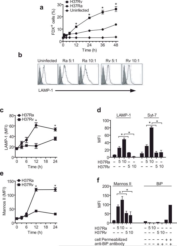
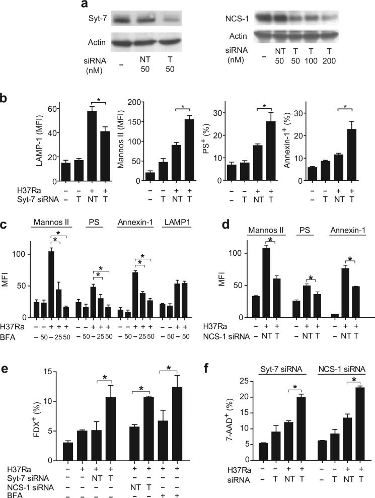
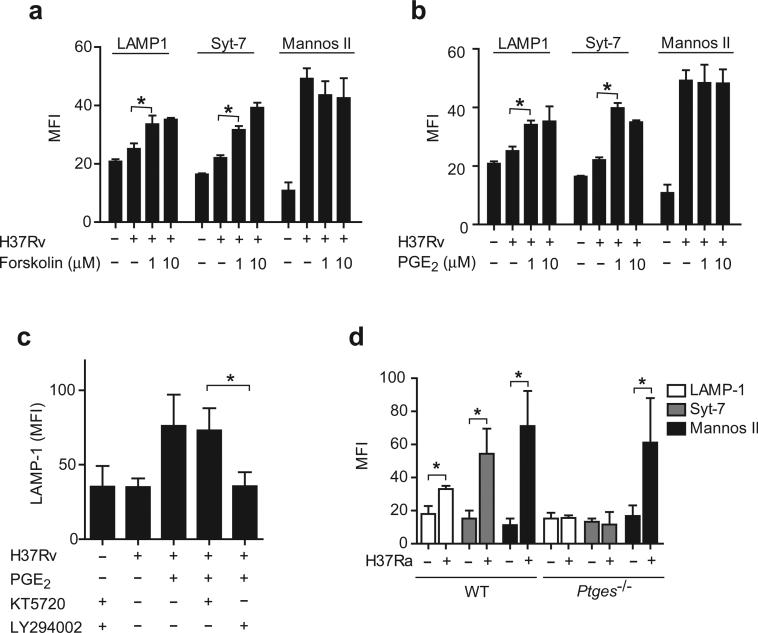
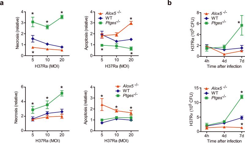
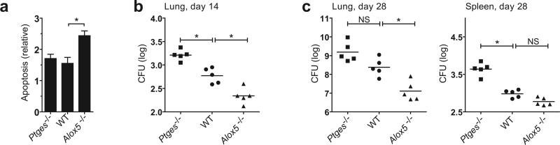
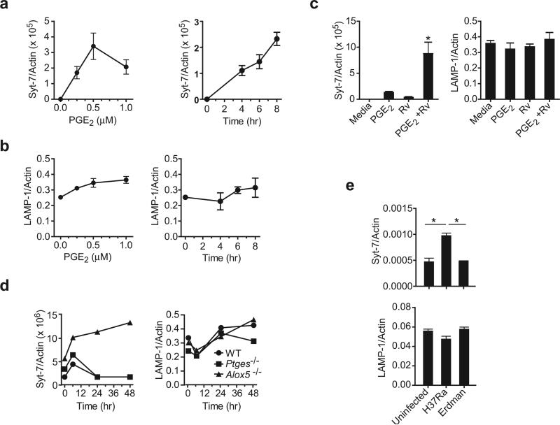
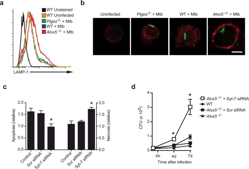
Induction of macrophage necrosis is a strategy used by virulent Mycobacterium tuberculosis (Mtb) to avoid innate host defense. In contrast, attenuated Mtb causes apoptosis, which limits bacterial replication and promotes T cell cross-priming by antigen-presenting cells. Here we show that Mtb infection causes plasma membrane microdisruptions. Resealing of these lesions, a process crucial for preventing necrosis and promoting apoptosis, required translocation of lysosomal and Golgi apparatus-derived vesicles to the plasma membrane. Plasma membrane repair depended on prostaglandin E(2) (PGE(2)), which regulates synaptotagmin 7 (Syt-7), the calcium sensor involved in the lysosome-mediated repair mechanism. By inducing production of lipoxin A(4) (LXA(4)), which blocks PGE(2) biosynthesis, virulent Mtb prevented membrane repair and induced necrosis. Thus, virulent Mtb impairs macrophage plasma membrane repair to evade host defenses.


Figures

Figure 1
Figure 1. Infection of human Mφ with virulent H37Rv inhibits lysosomal and Golgi-mediated plasma membrane repair
(a) Kinetics of FDX influx through membrane lesions in Mφ left uninfected or infected with H37Ra or H37Rv. (b) LAMP1 translocation to the plasma membrane lesions of human Mφ infected with H37Ra or H37Rv for 12 h. Shaded histogram is the isotype control and the open histogram is the specific LAMP1 staining. Numbers in histograms indicate the mean fluorescence intensity (MFI) of the entire cell population. Bacterial strain and MOI is indicated above each histogram. (c,e) Kinetics of LAMP1 (c) and mannosidase II (e) translocation to the surface of human Mφ infected with H37Ra or H37Rv. (d, f) Translocation of LAMP1, Syt-7, mannosidase II and the ER marker BiP to the surface of Mφ left uninfected or infected for 12 h with H37Ra or H37R. Where indicated, Mφ were permeabilized and stained with irrelevant (−) and BiP-specific (+) antibodies. The MOI was 5:1 (5) or 10:1 (10) where indicated; otherwise an MOI of 10:1 was used. Results in all panels are representative of at least three independent experiments (error bars, s.e.m.) *, p < 0.05.
Figure 2
Figure 2. Distinct Ca2+ sensors regulate recruitment of lysosome and Golgi apparatus derived membranes in Mtb infected human Mφ
(a) Expression of the Ca2+ sensors Syt-7 and NCS-1 after gene silencing in human Mφ (NT not targeted, T targeted) was measured by immunoblot. (b) Influence of Syt-7-specific siRNA on translocation of LAMP1, mannosidase II, PS, and annexin-1 to the surface of H37Ra-infected Mφ. (c) Influence of Brefeldin A (BFA) on translocation of LAMP1, mannosidase II, PS, and annexin-1 to the surface of H37Ra-infected Mφ. (d) Influence of NCS-1-specific siRNA on translocation of mannosidase II, PS, and annexin-1 to the surface of H37Ra-infected Mφ. (e) FDX influx into H37Ra infected Mφ expressing Syt-7 or NCS-1 specific siRNA or treated with Brefeldin A (50 uM). (f) Necrosis of H37Ra-infected Mφ expressing the indicated siRNA constructs. The MOI was 10:1. The results in each panel are from one representative of three independent experiments (error bars, s.e.m.) *, p < 0.05.
Figure 3
Figure 3. PGE2 reconstitutes lysosomal repair in human Mφ infected with virulent Mtb
(a,b) Translocation of LAMP1, Syt-7 and mannosidase II to the surface of H37Rv-infected Mφ (MOI of 10:1) treated with forskolin (1–10 μM) (a) or PGE2 (b). (c) LAMP1 translocation induced by H37Rv in presence of PGE2 (1 μM) after addition of the specific PI3K inhibitor (LY294002, 10 μM) and/or the PKA inhibitor (KT5720, 50 nM). (d), Translocation of Syt-7, LAMP1 and mannosidase II to the surface of H37Ra-infected wild-type (WT) and Ptges−/− Mφ. The results in each panel are from one representative of three independent experiments (error bars, s.e.m.) *, p < 0.05.
Figure 4
Figure 4. Bacterial growth and the death modality of Mtb infected murine Mφ is regulated by eicosanoids
(a) Apoptosis and necrosis three days after H37Rv (bottom) or H37Ra (top) infection of Alox5−/−, wild-type (WT) and Ptges−/− Mφ. Cell death was measured by ELISA. (b) Colony forming units (CFU) of H37Rv (bottom) or H37Ra (top) at indicated times after infection (MOI 10:1) of Alox5−/− Mφ, WT and Ptges−/− Mφ. Results are representative of 3 (a) and 2 (b) independent experiments (error bars, s.e.m.) *, p < 0.05.
Figure 5
Figure 5. The fate of Mtb-infected Mφ in vitro reflects the innate control of infection in vivo
(a) Alox5−/−, Ptges−/−, and wild-type (WT) mice (n=3 mice per group) were intratracheally nfected with H37Rv (1×10⁶ CFU) and BAL was collected 3 days after infection. Graph shows apoptosis of adherent antigen-presenting cells from infected Alox5−/−, WT and Ptges−/− mice as compared to uninfected controls. (b, c) Bacterial colony forming units in the spleen and/or lung 14 days (b) and 28 days (c) after intratracheal transfer of H37Rv infected Alox5−/−, Ptges−/−, or WT Mφ into Rag1−/− mice. Similar numbers of bacteria (WT log 10 = 1.81, Ptges−/− log 10 = 1.79, and Alox5−/− log 10 = 1.8) were found in the lungs of mice on day 1 after adoptive transfer. Data is from a single experiment with two time points (n=5 mice per group per time point); (error bars, s.e.m.) *, p < 0.05.
Figure 6
Figure 6. PGE2 regulates Syt-7 expression in murine Mφ
Dose and time response of (a) Syt-7 and (b) LAMP1 mRNA expression in naïve wild-type Mφ treated with PGE2. (c) Syt-7 and LAMP1 expression in wild-type Mφ infected or not with H37Rv after addition of PGE2 (1 μM). (d) Syt-7 and LAMP1 mRNA expression in Alox5−/−, Ptges−/− and wild-type (WT) Mφ at indicated times after infection with H37Rv. (e) Wild-type mice were infected or not by the aerosol route with a low dose (~ 100 CFU) of H37Rv or H37Ra. After 7 days of infection, RNA was extracted from the whole lung and the expression of Syt-7 and LAMP1 mRNA was measured by real-time PCR. The expression of Syt-7 or LAMP1 was normalized to β-Actin. Results are from one representative of 2 (a, b, c, and f) and 3 (d, e) independent experiments. n=3 mice per group; (error bars, s.e.m.) *, p < 0.05.
Figure 7
Figure 7. Syt-7 is essential for induction of plasma membrane repair, prevention of necrosis, and control of bacterial growth in murine Mφ
(a,b) Translocation of LAMP1 to the cell surface of Alox5−/−, Ptges−/−, and wild-type (WT) Mφ left uninfected or 24 h after infection with H37Rv (Mtb; MOI 5:1) as analyzed by flow cytometry (a) or confocal microscopy (b). In (b) Mφ were infected with GFP-labeled H37Rv (MOI 10:1), and nonpermeabilized cells were stained with a monoclonal antibody against the lumenal domain of LAMP1. Scale bar 5 μm. (c) Alox5−/− Mφ were left untreated or were transfected with scrambled (Scr) siRNA or siRNA specific for Syt-7 for 24 h followed by H37Rv infection (MOI 5:1). Apoptosis and necrosis of the treated and infected Mφ compared to uninfected controls was measured 3 days after infection. (d) H37Rv growth was measured in Alox5−/− and WT Mφ left untreated or transfected with Syt-7-specific or scrambled (Scr) siRNA at the indicated times after infection. Results are representative of 2 independent experiments. (error bars, s.e.m.) *, p < 0.05.

Similar articles

Cited by

References

    1. McNeil PL, Steinhardt RA. Plasma membrane disruption: repair, prevention, adaptation. Annu. Rev. Cell Dev. Biol. 2003;19:697–731. - PubMed
    1. Roy D, et al. A process for controlling intracellular bacterial infections induced by membrane injury. Science. 2004;304:1515–1518. - PubMed
    1. Smith J, et al. Evidence for pore formation in host cell membranes by ESX-1-secreted ESAT-6 and its role in Mycobacterium marinum escape from the vacuole. Infect. Immun. 2008;76:5478–5487. - PMC - PubMed
    1. Bi GQ, Alderton JM, Steinhardt RA. Calcium-regulated exocytosis is required for cell membrane resealing. J Cell Biol. 1995;131:1747–1758. - PMC - PubMed
    1. Togo T, Alderton JM, Bi GQ, Steinhardt RA. The mechanism of facilitated cell membrane resealing. J. Cell Sci. 1999;112(Pt 5):719–731. - PubMed

Publication types

MeSH terms

LinkOut - more resources