

Myopic traction maculopathy: study with spectral domain optical coherence tomography and review of the literature
- PMID: 19561782
- PMCID: PMC2683149
Myopic traction maculopathy: study with spectral domain optical coherence tomography and review of the literature
Abstract
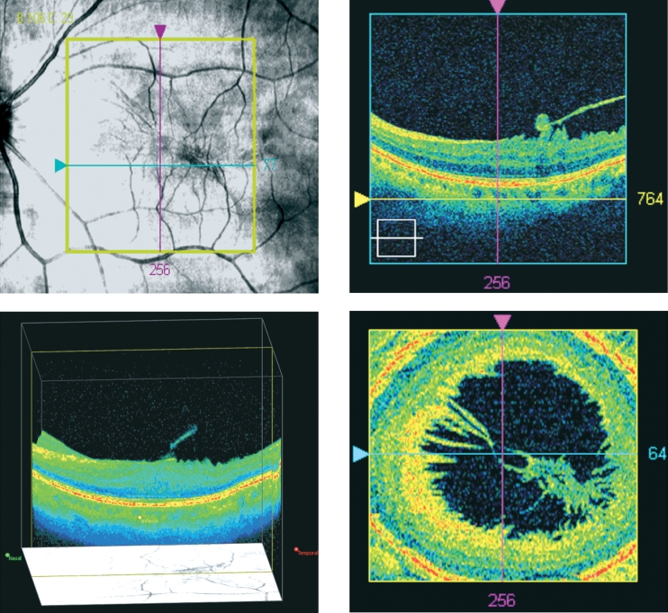
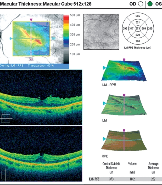
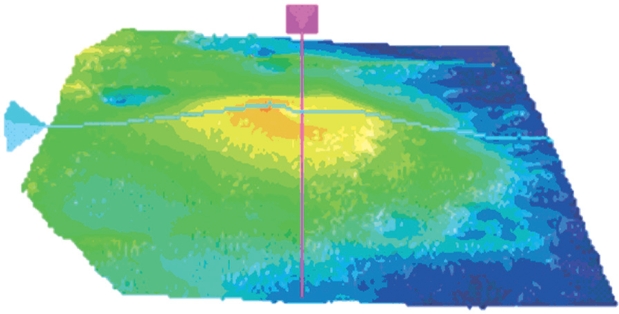
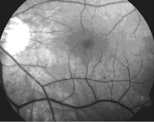
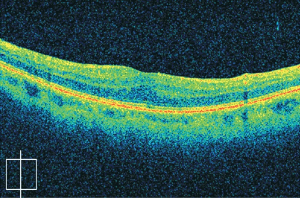
Aim: To describe the tomographic findings of a case of myopic traction maculopathy using Spectral Domain Optical Coherence Tomography (SD-OCT) and present the results of its surgical intervention.
Design: Observational case report and review of the literature.
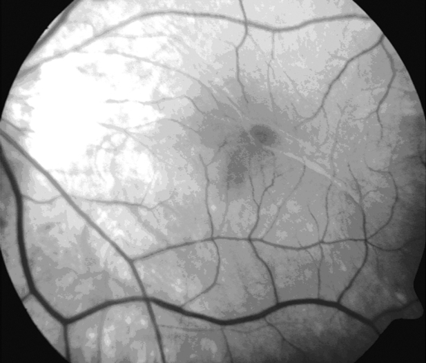
Methods: A 61-year-old male with metamorphopsia was examined clinically and with the use of SD-OCT. The diagnosis of myopic traction maculopathy was made. The patient underwent pars plana vitrectomy with removal of the vitreomacular adhesions, the epiretinal and the internal limiting membrane.
Results: Visual acuity increased by two Snellen lines, metamorphopsia disappeared, macular morphology was improved and myopic traction maculopathy was resolved.
Conclusions: Imaging with SD-OCT is capable of documentation and measurement of the early stages of myopic traction maculopathy. Moreover, vitrectomy was beneficial for the visual and anatomic outcome of the patient.
Keywords: myopic traction maculopathy; spectral domain optical coherence tomography.
Figures
References
-
- Saw SM, Katz J, Schein OD, et al. Epidemiology of myopia. Epidemiol Rev. 1996;18:175–187. - PubMed
-
- Curtin BJ, editor. The myopias: basic science and clinical management. Philadelphia: Harper and Row; 1985. pp. 7–10.
-
- Kanski JJ. Ch. 17, Degenerative myopia. In: Kanski JJ, editor. Clinical Ophthalmology, a systematic approach. 6th eds. Philadelphia: Elsevier; 2007. pp. 654–655.
-
- Takano M, Kishi S. Foveal retinoschisis and retinal detachment in severely myopic eyes with posterior staphyloma. Am J Ophthalmol. 1999;128:472–476. - PubMed
Publication types
LinkOut - more resources
Full Text Sources
Miscellaneous