. 2009 Jun 29;4(6):e6068.
doi: 10.1371/journal.pone.0006068.

A genetic screen in Drosophila reveals novel cytoprotective functions of the autophagy-lysosome pathway

Affiliations

A genetic screen in Drosophila reveals novel cytoprotective functions of the autophagy-lysosome pathway

Andrew M Arsham et al. PLoS One. .

Abstract

The highly conserved autophagy-lysosome pathway is the primary mechanism for breakdown and recycling of macromolecular and organellar cargo in the eukaryotic cell. Autophagy has recently been implicated in protection against cancer, neurodegeneration, and infection, and interest is increasing in additional roles of autophagy in human health, disease, and aging. To search for novel cytoprotective features of this pathway, we carried out a genetic mosaic screen for mutations causing increased lysosomal and/or autophagic activity in the Drosophila melanogaster larval fat body. By combining Drosophila genetics with live-cell imaging of the fluorescent dye LysoTracker Red and fixed-cell imaging of autophagy-specific fluorescent protein markers, the screen was designed to identify essential metazoan genes whose disruption causes increased flux through the autophagy-lysosome pathway. The screen identified a large number of genes associated with the protein synthesis and ER-secretory pathways (e.g. aminoacyl tRNA synthetases, Oligosaccharyl transferase, Sec61alpha), and with mitochondrial function and dynamics (e.g. Rieske iron-sulfur protein, Dynamin-related protein 1). We also observed that increased lysosomal and autophagic activity were consistently associated with decreased cell size. Our work demonstrates that disruption of the synthesis, transport, folding, or glycosylation of ER-targeted proteins at any of multiple steps leads to autophagy induction. In addition to illuminating cytoprotective features of autophagy in response to cellular damage, this screen establishes a genetic methodology for investigating cell biological phenotypes in live cells, in the context of viable wild type organisms.


Conflict of interest statement

Competing Interests: The authors have declared that no competing interests exist.

Figures

Figure 1
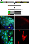
Figure 1. System of mitotic recombination for screening increased lysosomal activity in mosaic mutant Drosophila fat body.
Adult virgin females of the GFP indicator strain (y w hsflp; UAS-2xeGFP FRT40A Fb-Gal4, hereafter referred to as FRT40AGFP) were crossed to males carrying P-element-induced mutations distal to FRT40A on chromosome 2 L (A). 6–8 hr embryo collections were immediately heat shocked at 37°C for 1 hour to activate the Flp recombinase. Flp-catalyzed mitotic recombination and subsequent chromosomal segregation early in embryogenesis leads to three cellular genotypes, distinguishable in the larval fat body by their GFP dosage: mutant (−/−, 0 GFP); heterozygote (+/−, 1 GFP); and wild type (+/+, 2 GFP). (B) The top panels show a negative control cross between FRT40AGFP and an isogenized FRT40A wild type strain, with LysoTracker staining on the right. Bottom panels show mutant clones with markedly increased LysoTracker staining relative to neighboring internal control cells, a phenotype which we designate LT+. All panels are unfixed dissected tissue imaged epifluorescently.
Figure 2
Figure 2. Gene ontology categories for LT+ and LT− mutants.
Annotated genes associated with P-element insertions were assigned upper level gene ontology terms by the GO Term Mapper online software. The percentage of LT+ and LT− genes in selected categories are shown for cellular component (A), molecular function (B), and biological process (C). Not every gene is assigned GO terms, some genes have multiple GO terms, and categories are not mutually exclusive and therefore do not add up to 100%.
Figure 3
Figure 3. Disruption of tRNA synthetase genes increases LysoTracker staining.
(A) Clones of cells homozygous for P-element insertion KG03126 in the seryl tRNA synthetase CG17259 (indicated by white arrows and by lack of GFP in the inset panel) display punctate LysoTracker staining under fed conditions. (B and C) Cell clones expressing UAS-driven RNAi (indicated by GFP expression in inset panels) against phenylanalyl (B) and leucyl tRNA synthetases (C) display punctate LysoTracker staining. Allele used for mitotic clones: CG17259KG03126.
Figure 4
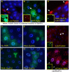
Figure 4. Disruption of mitochondria-associated genes leads to increased LysoTracker and autophagy.
Clones of cells homozygous for P-element insertions in ab, nmd, RISP, and Drp1 display punctate LysoTracker staining (A, D, F, H, unfixed tissue). Ab, nmd, and Drp1 mutations also lead to redistribution of fluorescent Atg8a into autophagosomes (B, E, I, fixed tissue, mutant clones are indicated here and elsewhere by solid white arrows; representative Atg8a punctae in B are indicated by arrowheads). Cells homozygous for the nmd insertion also show aberrant mitochondrial morphology as visualized by MitoTracker Red dye (C, fixed tissue) compared to neighboring cells. Cells expressing RNAi against RISP (indicated by GFP expression, G, unfixed tissue) have punctate and perinuclear LysoTracker staining. Inset panels indicate mutant clones which lack GFP or RFP, or Gal4-activated cells which express GFP, as appropriate. Alleles used for mitotic clones: abk02807, nmdk10909, RFeSPk11704, Drp1KG03815.
Figure 5
Figure 5. Disruption of the ER translocon induces autophagy.
Cells homozygous for P-element insertions in Sec61α display elevated and punctate LysoTracker (A) and GFP-Atg8a (B). Flies expressing RNAi against Sec61α also have elevated and punctate LysoTracker (C). (D–G) Flies expressing RNAi against Sec61α(Ε), Sec61βΦ, Sec61γ(Γ), have increased numbers of autophagosomes, compared to the no-RNAi control (D). Cells with elevated Sec61α suppress starvation-induced autophagy as measured by LysoTracker dye (H) and mCherry-Atg8a fusion protein (I). (A, C, H, unfixed tissue; B, D, E, F, G, I fixed tissue. Inset panels and arrows indicate mutant clones lacking GFP or RFP, or Gal4-activated cells which express GFP, as appropriate.) Allele used for mitotic clones: Sec61αk04917.
Figure 6
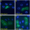
Figure 6. Disruption of the signal recognition particle and its receptor induce autophagosome formation.
Cells expressing inducible RNAi against the indicated subunits of the SRP and SRP receptor, marked by GFP-Atg8a expression, have elevated numbers of autophagosomes as indicated by punctate localization of GFP fluorescence. A, B, C, D, fixed tissue.
Figure 7
Figure 7. Disruption of a putative GlcNAc-1 phosphotransferase (CG5287) and the homolog of mammalian oligosaccharyl transferase (CG13393) cause increased LysoTracker staining.
Mitotic mutant clones for P-element insertions in the genes CG5287 (A) and CG13393 (B) have elevated punctate LysoTracker staining. Inset: GFP expression identifies mutant clones (white arrows). Alleles used for mitotic clones: CG5287k10105, CG13393k12914.
Figure 8
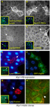
Figure 8. Involvement of the unfolded protein response in lysosomal expansion and autophagy.
Fat body cells mutant for Sec61α (A) or expressing RNAi against Sec61γ or SrpRβ (B and C) and immunostained with antibody to Hsc70–3 have elevated levels of the protein relative to neighboring wild type cells, or cells expressing GFP alone (D). Inset panels and white arrows indicate cells of interest. Cells expressing UAS-driven constitutively active Xbp1-RB display punctate LysoTracker (E) and autophagosome formation (F; representative punctae indicated by arrowheads). Cells homozygous for a P-element insertion in Xbp1 display both elevated LysoTracker (G) and autophagosome formation (H). Genotypes used for mitotic clone formation: y w hsflp; Cg-GAL4 FRT42D UAS-GFP (G) or y w hsflp myr-dsRed FRT42D GFP-Atg8a (H), y w; FRT42D P{lacW}Xbp1k13803/SM6-TM6B. A, B, C, D, F, H, fixed tissue; E, G, unfixed tissue.

References

    1. Ravid T, Hochstrasser M. Diversity of degradation signals in the ubiquitin-proteasome system. Nat Rev Mol Cell Biol. 2008;9:679–690. - PMC - PubMed
    1. Xie Z, Klionsky DJ. Autophagosome formation: core machinery and adaptations. Nat Cell Biol. 2007;9:1102–1109. - PubMed
    1. Vellodi A. Lysosomal storage disorders. Br J Haematol. 2005;128:413–431. - PubMed
    1. Mizushima N, Levine B, Cuervo AM, Klionsky DJ. Autophagy fights disease through cellular self-digestion. Nature. 2008;451:1069–1075. - PMC - PubMed
    1. Tsukada M, Ohsumi Y. Isolation and characterization of autophagy-defective mutants of Saccharomyces cerevisiae. FEBS Lett. 1993;333:169–174. - PubMed

Publication types

Substances