

Systems approach to explore components and interactions in the presynapse
- PMID: 19562802
- PMCID: PMC2766278
- DOI: 10.1002/pmic.200800767
Systems approach to explore components and interactions in the presynapse
Abstract
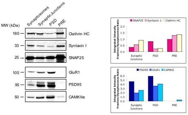
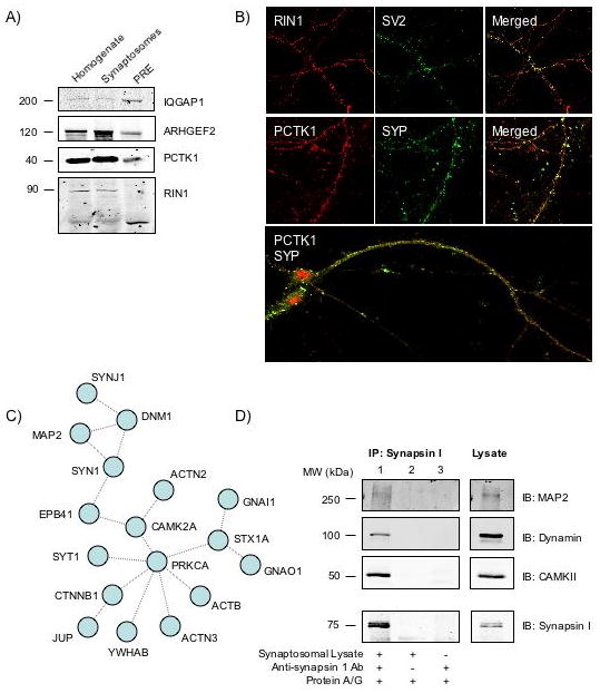
The application of proteomic techniques to neuroscientific research provides an opportunity for a greater understanding of nervous system structure and function. As increasing amounts of neuroproteomic data become available, it is necessary to formulate methods to integrate these data in a meaningful way to obtain a more comprehensive picture of neuronal subcompartments. Furthermore, computational methods can be used to make biologically relevant predictions from large proteomic data sets. Here, we applied an integrated proteomics and systems biology approach to characterize the presynaptic (PRE) nerve terminal. For this, we carried out proteomic analyses of presynaptically enriched fractions, and generated a PRE literature-based protein-protein interaction network. We combined these with other proteomic analyses to generate a core list of 117 PRE proteins, and used graph theory-inspired algorithms to predict 92 additional components and a PRE complex containing 17 proteins. Some of these predictions were validated experimentally, indicating that the computational analyses can identify novel proteins and complexes in a subcellular compartment. We conclude that the combination of techniques (proteomics, data integration, and computational analyses) used in this study are useful in obtaining a comprehensive understanding of functional components, especially low-abundance entities and/or interactions in the PRE nerve terminal.
Conflict of interest statement
The authors have no conflicts of interest to declare.
Figures

References
-
- Abul-Husn NS, Devi LA. Neuroproteomics of the synapse and drug addiction. J Pharmacol Exp Ther. 2006;318:461–468. - PubMed
-
- Schrimpf SP, Meskenaite V, Brunner E, Rutishauser D, et al. Proteomic analysis of synaptosomes using isotope-coded affinity tags and mass spectrometry. Proteomics. 2005;5:2531–2541. - PubMed
Publication types
MeSH terms
Substances
Grants and funding
- K02 DA000458/DA/NIDA NIH HHS/United States
- K05 DA019521/DA/NIDA NIH HHS/United States
- R01 NS026880/NS/NINDS NIH HHS/United States
- DA08863/DA/NIDA NIH HHS/United States
- RR017802/RR/NCRR NIH HHS/United States
- S10 RR022415/RR/NCRR NIH HHS/United States
- S10 RR017802/RR/NCRR NIH HHS/United States
- R24 CA088325/CA/NCI NIH HHS/United States
- K04 NS001788/NS/NINDS NIH HHS/United States
- R01 DA008863/DA/NIDA NIH HHS/United States
- R03 NS053751/NS/NINDS NIH HHS/United States
- R56 DA008863/DA/NIDA NIH HHS/United States
- 1P50GM071558-01A27398/GM/NIGMS NIH HHS/United States
- R13 DA025367/DA/NIDA NIH HHS/United States
- R01 DK038761/DK/NIDDK NIH HHS/United States
- P50 GM071558/GM/NIGMS NIH HHS/United States
- R37 DA008863/DA/NIDA NIH HHS/United States
- P30 NS061777/NS/NINDS NIH HHS/United States
- GM54508/GM/NIGMS NIH HHS/United States
- T32 DA007135/DA/NIDA NIH HHS/United States
- P41 RR000862/RR/NCRR NIH HHS/United States
- R13 DA026655/DA/NIDA NIH HHS/United States
- R24 CA095823/CA/NCI NIH HHS/United States
- DK 38761/DK/NIDDK NIH HHS/United States
- R01 GM054508/GM/NIGMS NIH HHS/United States
- CA88325/CA/NCI NIH HHS/United States
- DA019521/DA/NIDA NIH HHS/United States
LinkOut - more resources
Full Text Sources
Molecular Biology Databases
