Review
. 2009 Jul;19(3):483-92.
doi: 10.1111/j.1750-3639.2009.00289.x.

Myofibrillar myopathies: a clinical and myopathological guide

Affiliations
Review

Myofibrillar myopathies: a clinical and myopathological guide

Rolf Schröder et al. Brain Pathol. 2009 Jul.

Abstract

Myofibrillar myopathies (MFMs) are histopathologically characterized by desmin-positive protein aggregates and myofibrillar degeneration. Because of the marked phenotypic and pathomorphological variability, establishing the diagnosis of MFM can be a challenging task. While MFMs are partly caused by mutations in genes encoding for extramyofibrillar proteins (desmin, alphaB-crystallin, plectin) or myofibrillar proteins (myotilin, Z-band alternatively spliced PDZ-containing protein, filamin C, Bcl-2-associated athanogene-3, four-and-a-half LIM domain 1), a large number of these diseases are caused by still unresolved gene defects. Although recent years have brought new insight into the pathogenesis of MFMs, the precise molecular pathways and sequential steps that lead from an individual gene defect to progressive muscle damage are still unclear. This review focuses on the clinical and myopathological aspects of genetically defined MFMs, and shall provide a diagnostic guide for this numerically significant group of protein aggregate myopathies.


Figures

Figure 1
Figure 1
Schematic drawing of a muscle fiber with regard to myofibrillar myopathies (MFMs) causing mutations in genes coding for extramyofibrillar (desmin, αB‐crystallin, plectin) and myofibrillar (filamin C, myotilin, Z‐band alternatively spliced PDZ‐containing protein, four‐and‐a‐half LIM domain 1, Bcl‐2‐associated athanogene‐3) proteins. Note that the extramyofibrillar cytoskeleton forms a three‐dimensional scaffold around myofibrillar Z‐disks, thereby interlinking neighboring myofibrils and connecting the myofibrillar apparatus to myonuclei, sarcolemma and mitochondria. Mutations in these known MFM genes lead to misfolded proteins and desmin‐positive protein aggregates in conjunction with Z‐disk alterations and mitochondrial abnormalities. The ubiquitin–proteasome and the autophagic–lysosomal system are essential components of the cellular protein quality control framework, which is responsible for the degradation of misfolded proteins and protein aggregates.
Figure 2
Figure 2
Histopathological findings in myofibrillar myopathies (MFMs). Hematoxylin & eosin (H&E) (A) and Gomori trichrome (G‐Tri) (B) staining in a genetically proven desminopathy. Arrows indicate the presence of isolated sarcoplasmic and subsarcolemmal protein aggregates (bars = 50 µm). H&E (C) and G‐Tri (D) staining in a genetically proven myotilinopathy and in a patient with MFM of unknown aetiology, respectively. Note the vacuolar changes (arrows) in myotilinopathy (bar = 75 µm) and the polymorphic protein aggregates in MFM of unknown aetiology (bar = 50 µm). Succinic dehydrogenase (E) and cytochrome‐C oxidase (F) staining in a genetically proven desminopathy. Note the presence of rubbed‐out fibers (*) and multiple core‐like lesions (bars = 40 µm). Bar in (A), (B), (D) = 50 µm. Bar in (C) = 75 µm. Bar in (E), (F) = 40 µm.
Figure 3
Figure 3
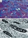
Spheroid bodies (arrows) appear as coiled aggregates arranged in linear packets of greenish material in Gomori trichrome stain (A). At the ultrastructural level (B), a spheroid body is a well‐circumscribed and demarcated body (asterisk). This body is devoid of mitochondria or other cytoplasmic organelles. A spheroid body is highly indicative of myotilinopathy. Bar in (A) = 30 µm.
Figure 4
Figure 4
Reducing bodies are characterized by the presence of intracytoplasmic inclusion bodies strongly dark blue stained in the menadione‐linked α‐glycerophosphate dehydrogenase preparation without substrate, α‐glycerophosphate. Here (A), an Azan stain nicely reveals numerous reducing bodies in many muscle fibers (arrows). Electron microscopy (B) shows reducing bodies (asterisk) composed of dense osmiophilic material consisting of closely packed granulofibrillar particles. Myofibrils with reducing bodies have disorganized cross‐striations. This condition is also commonly associated with rimmed vacuoles and cytoplasmic bodies. A reducing body is highly indicative of FHL1opathy. Bar in (A) = 40 µm.
Figure 5
Figure 5
Myopathological findings in an epidermolysis bullosa simplex with muscular dystrophy patient with a homozygous plectin mutation (31). Hematoxylin & eosin (A) and cytochrome‐C oxidase (COX) (B) stains demonstrate severe myopathic and mitochondrial alternations. * denotes a COX‐negative fiber. (C) Note the subsarcolemmal and sarcoplasmic accumulation of desmin‐positive material. (D) Desmin immunogold electron microscopy shows highly unordered desmin filaments labeled by multiple gold particles. * denotes cross‐sectioned myofibrils. Bar in (A) = 50 µm.
Figure 6
Figure 6
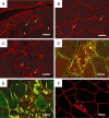
Indirect immunofluorescence findings in myofibrillar myopathies. Desmin (A–C) staining in genetically proven desminopathies. Note the high variability of protein aggregate formation. Arrows in (A) denote sarcoplasmic and arrowheads subsarcolemmal aggregates. In (B), desmin‐positive aggregates are restriced to the subsarcolemmal region. An extensive vacuolar presentation of a desminopathy is demonstrated in (C). Desmin‐ and αB‐crystallin‐positive inclusions in genetically proven myotilinopathy and desminopathy are highlighted in (D) and (E), respectively. (F) Caveolin‐3‐positive membranous structures within muscle fibers of a genetically proven desminopathy. Bar in (A) = 60 µm. Bar in (C) = 100 µm. Bar in (B), (D), (E), (F) = 50 µm.
Figure 7
Figure 7
Electron microscopy findings in myofibrillar myopathy. (A,B) Granulofilamentous material (*) in genetically proven desminopathy. Note the clustering of mitochondria (arrow) in close relation to granulofilamentous material and myofibrillar structures. (C,D) Filamentous sarcoplasmic inclusions (*) in genetically proven filaminopathy. Arrows denote Z‐disk remnants.

References

    1. Bär H, Fischer D, Goudeau B, Kley RA, Clemen CS, Vicart P et al (2005) Pathogenic effects of a novel heterozygous r350p desmin mutation on the assembly of desmin intermediate filaments in vivo and in vitro . Hum Mol Genet 14:1251–1260. - PubMed
    1. Bär H, Mücke N, Kostareva A, Sjöberg G, Aebi U, Herrmann H (2005) Severe muscle disease‐causing desmin mutations interfere with in vitro filament assembly at distinct stages. Proc Natl Acad Sci U S A 102:15099–115104. - PMC - PubMed
    1. Bär H, Kostareva A, Sjöberg G, Sejersen T, Katus HA, Herrmann H (2006) Forced expression of desmin and desmin mutants in cultured cells: impact of myopathic missense mutations in the central coiled‐coil domain on network formation. Exp Cell Res 312:1554–1565. - PubMed
    1. Bär H, Goudeau B, Walde S, Casteras‐Simon M, Mücke N, Shatunov A et al (2007) Conspicuous involvement of desmin tail mutations in diverse cardiac and skeletal myopathies. Hum Mutat 28:374–386. - PubMed
    1. Bär H, Mücke N, Katus HA, Aebi U, Herrmann H (2007) Assembly defects of desmin disease mutants carrying deletions in the alpha‐helical rod domain are rescued by wild type protein. J Struct Biol 158:107–115. - PubMed

Publication types