

Vascular dysfunction in the diabetic placenta: causes and consequences
- PMID: 19563553
- PMCID: PMC2714640
- DOI: 10.1111/j.1469-7580.2009.01098.x
Vascular dysfunction in the diabetic placenta: causes and consequences
Abstract
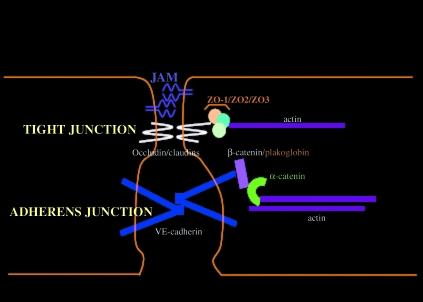
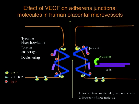
The development and functioning of the human fetoplacental vascular system are vulnerable to the maternal diabetic milieu. These vessels are in direct continuum with the fetal vascular system and are therefore also vulnerable to fetal endocrine derangements. Increased angiogenesis, altered junctional maturity and molecular occupancy, together with increased leakiness, constitute a well-described phenotype of vessels in the Type 1 diabetic human placenta and can be related to increased levels of placental vascular endothelial growth factor. The causes of these observed changes, whether maternal hyperglycaemia or fetal hyperinsulinaemia, still remain to be shown in the human placenta. Mechanistic studies using different vascular systems have shown high glucose and insulin to have profound vascular effects, with elevations in vascular endothelial growth factor, nitric oxide and protein kinase C being behind alterations in junctional adhesion molecules such as occludin and vascular endothelial-cadherin and vascular leakage of albumin. The role of advanced glycation products and oxidative stress in this vascular pathology is also discussed. The altered molecular mechanisms underlying the vascular changes in the diabetic human placenta may reflect similar consequences of high glucose and hyperinsulinaemia.
Figures
Similar articles
-
Vascular endothelial cadherin and beta-catenin in human fetoplacental vessels of pregnancies complicated by Type 1 diabetes: associations with angiogenesis and perturbed barrier function.Diabetologia. 2004 Apr;47(4):695-709. doi: 10.1007/s00125-004-1341-7. Diabetologia. 2004. PMID: 15298347
-
Placental vascular dysfunction in diabetic pregnancies: intimations of fetal cardiovascular disease?Microcirculation. 2011 May;18(4):263-9. doi: 10.1111/j.1549-8719.2011.00091.x. Microcirculation. 2011. PMID: 21418381 Review.
-
Enhanced fetoplacental angiogenesis in pre-gestational diabetes mellitus: the extra growth is exclusively longitudinal and not accompanied by microvascular remodelling.Diabetologia. 2002 Oct;45(10):1434-9. doi: 10.1007/s00125-002-0927-1. Epub 2002 Sep 5. Diabetologia. 2002. PMID: 12378385
-
Oxidative stress causes vascular dysfunction in the placenta.J Matern Fetal Med. 2000 Jan-Feb;9(1):79-82. doi: 10.1002/(SICI)1520-6661(200001/02)9:1<79::AID-MFM16>3.0.CO;2-O. J Matern Fetal Med. 2000. PMID: 10757441 Review.
-
Insulin action on the human placental endothelium in normal and diabetic pregnancy.Curr Vasc Pharmacol. 2009 Oct;7(4):460-6. doi: 10.2174/157016109789043973. Curr Vasc Pharmacol. 2009. PMID: 19485889 Review.
Cited by
-
Low molecular weight heparin (nadroparin) improves placental permeability in rats with gestational diabetes mellitus via reduction of tight junction factors.Mol Med Rep. 2020 Feb;21(2):623-630. doi: 10.3892/mmr.2019.10868. Epub 2019 Dec 6. Mol Med Rep. 2020. PMID: 31974593 Free PMC article.
-
The Placental Role in Gestational Diabetes Mellitus: A Molecular Perspective.touchREV Endocrinol. 2024 Apr;20(1):10-18. doi: 10.17925/EE.2024.20.1.5. Epub 2024 Mar 14. touchREV Endocrinol. 2024. PMID: 38812661 Free PMC article. Review.
-
Pregnancy in women with diabetic nephropathy.J Nephrol. 2019 Jun;32(3):379-388. doi: 10.1007/s40620-018-0553-8. Epub 2018 Nov 15. J Nephrol. 2019. PMID: 30430413 Review.
-
Adiponectin exerts antiproliferative effect on human placenta via modulation of the JNK/c-Jun pathway.Int J Clin Exp Pathol. 2014 May 15;7(6):2894-904. eCollection 2014. Int J Clin Exp Pathol. 2014. PMID: 25031708 Free PMC article.
-
Placental maternally expressed gene 3 differentially methylated region methylation profile is associated with maternal glucose concentration and newborn birthweight.J Diabetes Investig. 2021 Jun;12(6):1074-1082. doi: 10.1111/jdi.13432. Epub 2020 Nov 4. J Diabetes Investig. 2021. PMID: 33090678 Free PMC article.
References
-
- Aiello LP, Avery RL, Arrigg PG, et al. Vascular endothelial growth factor in ocular fluid of patients with diabetic retinopathy and other retinal disorders. N Engl J Med. 1994;331:1480–1487. - PubMed
-
- Antonetti DA, Barber AJ, Khin SE, et al. Vascular permeability in experimental diabetes is associated with reduced endothelial occludin content. Vascular endothelial growth factor decreases occludin in retinal endothelial cells. Diabetes. 1998;47:1953–1959. - PubMed
-
- Babawale MO, Lovat S, Mayhew TM, Lammiman MJ, James DK, Leach L. Effects of gestational diabetes on junctional adhesion molecules in human term placenta vasculature. Diabetologia. 2000;43:1185–1196. - PubMed
-
- Bazzoni G, Dejana E. Endothelial cell-to-cell junctions: molecular organization and role in vascular homeostasis. Physiol Rev. 2003;84:869–901. - PubMed
-
- Ben-Ze’ev A, Geiger B. Differential molecular interactions of β-catenin and plakoglobin in adhesion, signalling and cancer. Curr Opin Cell Biol. 1998;10:629–639. - PubMed
Publication types
MeSH terms
Grants and funding
LinkOut - more resources
Full Text Sources
Medical
