Comparative Study
. 2009 Jun 29:9:59.
doi: 10.1186/1472-6750-9-59.

Novel sequential ChIP and simplified basic ChIP protocols for promoter co-occupancy and target gene identification in human embryonic stem cells

Affiliations
Comparative Study

Novel sequential ChIP and simplified basic ChIP protocols for promoter co-occupancy and target gene identification in human embryonic stem cells

Ricardo B Medeiros et al. BMC Biotechnol. .

Abstract

Background: The investigation of molecular mechanisms underlying transcriptional regulation, particularly in embryonic stem cells, has received increasing attention and involves the systematic identification of target genes and the analysis of promoter co-occupancy. High-throughput approaches based on chromatin immunoprecipitation (ChIP) have been widely used for this purpose. However, these approaches remain time-consuming, expensive, labor-intensive, involve multiple steps, and require complex statistical analysis. Advances in this field will greatly benefit from the development and use of simple, fast, sensitive and straightforward ChIP assay and analysis methodologies.

Results: We initially developed a simplified, basic ChIP protocol that combines simplicity, speed and sensitivity. ChIP analysis by real-time PCR was compared to analysis by densitometry with the ImageJ software. This protocol allowed the rapid identification of known target genes for SOX2, NANOG, OCT3/4, SOX17, KLF4, RUNX2, OLIG2, SMAD2/3, BMI-1, and c-MYC in a human embryonic stem cell line. We then developed a novel Sequential ChIP protocol to investigate in vivo promoter co-occupancy, which is basically characterized by the absence of antibody-antigen disruption during the assay. It combines centrifugation of agarose beads and magnetic separation. Using this Sequential ChIP protocol we found that c-MYC associates with the SOX2/NANOG/OCT3/4 complex and identified a novel RUNX2/BMI-1/SMAD2/3 complex in BG01V cells. These two TF complexes associate with two distinct sets of target genes. The RUNX2/BMI-1/SMAD2/3 complex is associated predominantly with genes not expressed in undifferentiated BG01V cells, consistent with the reported role of those TFs as transcriptional repressors.

Conclusion: These simplified basic ChIP and novel Sequential ChIP protocols were successfully tested with a variety of antibodies with human embryonic stem cells, generated a number of novel observations for future studies and might be useful for high-throughput ChIP-based assays.


Figures

Figure 1
Figure 1
Simplified, basic ChIP protocol schematic representation.
Figure 2
Figure 2
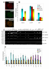
Identification of SOX2/NANOG/OCT3/4 target genes using the simplified, basic ChIP protocol. (A) NANOG expression by immunocytochemistry (red), DAPI (green) and merged images of undifferentiated BG01V human embryonic stem cells (magnification = 20×). (B) rtPCR data from SOX2/NANOG/OCT3/4 ChIP using primers to previously known target genes. (C) PCR results from the indicated ChIP assays with undifferentiated BG01V cells. (D) Densitometric analysis of ChIP data shown in C, obtained with ImageJ software. Three independent experiments were performed. Representative results are shown. Fold enrichment = ChIP/Input DNA. Error bars represent standard deviation.
Figure 3
Figure 3
Identification of known target genes for a variety of stem cell transcription factors using the simplified, basic ChIP protocol. (A) Mouse Lama1 detection from SOX17 ChIP using endoderm differentiated D3 mouse embryonic stem cells. (B) VEGF and BAX detection from RUNX2 ChIP. (C) OCT3/4, NANOG, and B2R detection from KLF4 ChIP. (D) HOXC13 detection from BMI-1 ChIP. (E) c-MYC and GLI1 detection from SMAD2/3 ChIP. (F) P21 detection from OLIG2 ChIP. (A) - (F): Fold enrichment = ChIP/Input DNA. Error bars represent standard deviation. (B-F): using undifferentiated BG01V human embryonic stem cells.
Figure 4
Figure 4
Specificity and applicability of the simplified, basic ChIP protocol. (A) As an additional negative control, the insulin promoter could not be detected from PDX1 ChIP assays using undifferentiated BG01V human embryonic stem cells. (B) The simplified, basic ChIP protocol (ChIP 1) was directly compared to previously described [20] commonly used ChIP protocol (ChIP 2), using three different antibodies (SOX17, BMI-1, OLIG2) and their respective known target genes (Lama1, HOXC13, p21). (C) ImageJ measurements (mean gray values) of PCR products from OLIG2 ChIP assays (p21 promoter detection), using a serial dilutions of DNA sample (from OLIG2 ChIP) and p21 promoter primers (logarithmic curve was added to the bar graphic). Fold enrichment = ChIP/Input DNA. Error bars represent standard deviation.
Figure 5
Figure 5
Sequential ChIP assay schematic representation.
Figure 6
Figure 6
Identification of SOX2/OCT3/4/NANOG/c-MYC and RUNX2/BMI-1/SMAD2/3 as complexes in BG01V cells using the novel Sequential ChIP protocol. (A) Detection of promoter co-occupancy by SOX2/OCT3/4/NANOG, with the indicated combinations of antibodies. (B) Detection of promoter co-occupancy by SOX2/OCT3/4/NANOG/c-MYC, with the indicated combinations of antibodies. (C) SOX2 immunodepletion of BG01V lysates by three subsequent rounds of SOX2 immumoprecipitation, using ERK1/2 as a negative control. (D) c-MYC ChIP assays results using SOX2-depleted or non-depleted BG01V human embryonic stem cell lysates. (E) Detection of promoter co-occupancy by BMI-1/RUNX2/SMAD2/3, with the indicated combinations of antibodies.

References

    1. Levine M, Tjian R. Transcription regulation and animal diversity. Nature. 2003;424:147–151. doi: 10.1038/nature01763. - DOI - PubMed
    1. Meng X, Brodsky MH, Wolfe SA. A bacterial one-hybrid system for determining the DNA-binding specificity of transcription factors. Nat Biotechnol. 2005;23:988–994. doi: 10.1038/nbt1120. - DOI - PMC - PubMed
    1. Mukherjee S, Berger MF, Jona G, Wang XS, Muzzey D, Snyder M, Young RA, Bulyk ML. Rapid analysis of the DNA-binding specificities of transcription factors with DNA microarrays. Nat Genet. 2004;36:1331–1339. doi: 10.1038/ng1473. - DOI - PMC - PubMed
    1. Walhout AJ. Unraveling transcription regulatory networks by protein-DNA and protein-protein interaction mapping. Genome Res. 2006;16:1445–1454. doi: 10.1101/gr.5321506. - DOI - PubMed
    1. Solomon MJ, Larsen PL, Varshavsky A. Mapping protein-DNA interactions in vivo with formaldehyde: evidence that histone H4 is retained on a highly transcribed gene. Cell. 1988;53:937–947. doi: 10.1016/S0092-8674(88)90469-2. - DOI - PubMed

Publication types