. 2009 Jun 26;137(7):1213-24.
doi: 10.1016/j.cell.2009.03.049.

Structure of N-terminal domain of NPC1 reveals distinct subdomains for binding and transfer of cholesterol

Affiliations

Structure of N-terminal domain of NPC1 reveals distinct subdomains for binding and transfer of cholesterol

Hyock Joo Kwon et al. Cell. .

Abstract

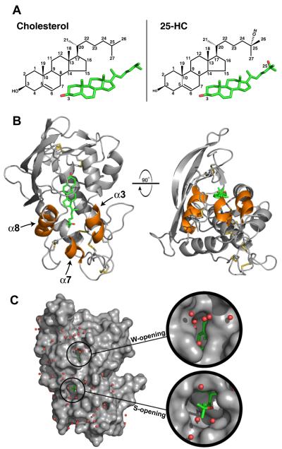
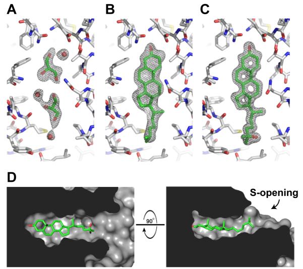
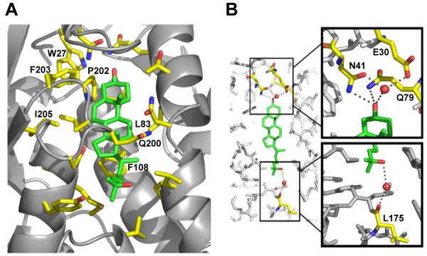
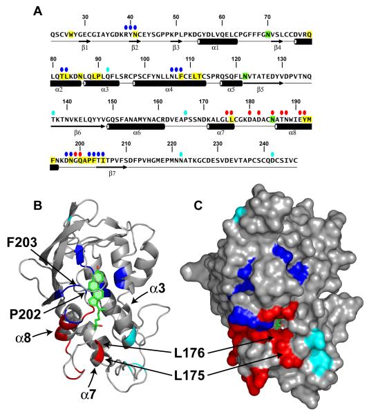
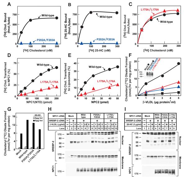
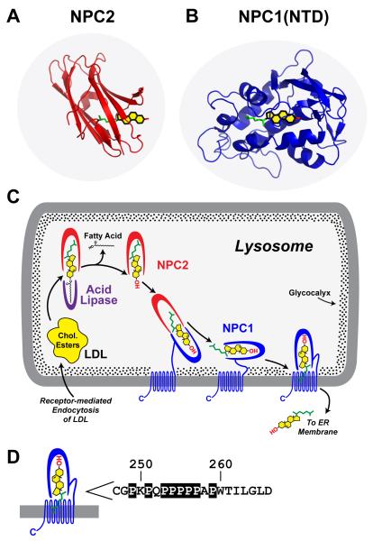
LDL delivers cholesterol to lysosomes by receptor-mediated endocytosis. Exit of cholesterol from lysosomes requires two proteins, membrane-bound Niemann-Pick C1 (NPC1) and soluble NPC2. NPC2 binds cholesterol with its isooctyl side chain buried and its 3beta-hydroxyl exposed. Here, we describe high-resolution structures of the N-terminal domain (NTD) of NPC1 and complexes with cholesterol and 25-hydroxycholesterol. NPC1(NTD) binds cholesterol in an orientation opposite to NPC2: 3beta-hydroxyl buried and isooctyl side chain exposed. Cholesterol transfer from NPC2 to NPC1(NTD) requires reorientation of a helical subdomain in NPC1(NTD), enlarging the opening for cholesterol entry. NPC1 with point mutations in this subdomain (distinct from the binding subdomain) cannot accept cholesterol from NPC2 and cannot restore cholesterol exit from lysosomes in NPC1-deficient cells. We propose a working model wherein after lysosomal hydrolysis of LDL-cholesteryl esters, cholesterol binds NPC2, which transfers it to NPC1(NTD), reversing its orientation and allowing insertion of its isooctyl side chain into the outer lysosomal membranes.


Figures

Figure 1
Figure 1. Structure of NPC1(NTD) Bound to 25-Hydroxycholesterol
(A) Stick models of cholesterol (left) and 25-HC (right) with carbon positions numbered. Carbon and oxygen atoms are colored green and red, respectively. (B) NPC1(NTD) is represented as a ribbon diagram in gray, and the disulfide bonds are shown in yellow. The positions of cholesterol and 25-HC are essentially identical. For simplicity, we show only the 25-HC molecule (colored in green). Helix3, helix7, and helix8 are colored orange. (C) The surface of NPC1(NTD), colored in gray, reveals openings at either end of the bound sterol. The W-opening would allow passage of a single water molecule (red spheres), but not a sterol molecule. The S-opening would become large enough to allow entry or exit of a sterol if the opening were expanded slightly (see Figure 2D).
Figure 2
Figure 2. Sterol Binding Pocket
(A-C) The sigmaA-weighted 2Fo-Fc electron density map contoured at 1σ within the sterol binding pocket is shown in gray mesh for the apoNPC1(NTD) (A), the cholesterol-bound (B), and 25-HC-bound (C) forms. The binding pocket is essentially identical in apoNPC1(NTD) and the sterol bound forms. The binding pocket of apoNPC1(NTD) is occupied by 2 glycerol and 3 water molecules. NPC1(NTD) is colored gray, bound ligands are colored green, and water molecules are shown as red spheres. (D) The internal surface of the binding pocket is shown in gray. Bound 25-HC is shown in green.
Figure 3
Figure 3. Expanded View of Sterol Binding Pocket
(A) Residues that line the binding pocket are shown in yellow, and the sterol molecule is shown in green. (B) Hydrogen bonds between residues of NPC1(NTD) and the hydroxyl groups of the sterol are denoted by gray dots. Residues involved in hydrogen bonds are colored yellow, and the sterol molecule is colored green. Nitrogen atoms are shown in blue, and oxygen atoms are shown in red. Water molecules are shown as red spheres.
Figure 4
Figure 4. Location of Functionally Important Residues in NPC1(NTD)
(A) Amino acid sequence of NPC1(NTD) with functionally important residues highlighted. Blue ovals denote residues that exhibit decreased binding of cholesterol and 25-HC by >75% when mutated to alanine. Red ovals denote residues that exhibit decreased transfer of cholesterol to liposomes by >70% when mutated to alanine. Cyan ovals denote naturally-occurring mutations in patients with NPC1 disease. Residues that line the binding pocket are shaded yellow. N-linked glycosylation sites that were eliminated are shaded green. The secondary structure of NPC1(NTD) is indicated below the sequence. (B and C) Ribbon diagram (B) and surface representation (C) of NPC1(NTD), showing the positions of functionally important residues. Bound 25-HC is shown as a stick model in green. Color coding is the same as in (A). The location of the L175, L176, P202, and F203 residues are denoted by arrows.
Figure 5
Figure 5. Biochemical and Functional Analysis of Sterol Binding and Transfer Mutants
(A-C) 3H-Sterol binding. Each reaction, in a final volume of 80 μl buffer C with 0.004% NP-40, contained 220 ng purified WT or mutant NPC1(NTD)-LVPRGS-His8-FLAG, 1 μg BSA, and indicated concentration of [3H]cholesterol (132 dpm/fmol) (A,C) or [3H]25-HC (165 dpm/fmol) (B). After incubation for 24 hr at 4°C, the amount of bound 3H-sterol was measured as described in Supplemental Experimental Procedures. Each value is the average of duplicate assays and represents total binding after subtraction of a blank value (10-70 fmol/tube). Mean variation for each of the duplicate assays in A, B, and C were 6.1%, 5.0%, and 5.0%, respectively. (D) [3H]Cholesterol transfer from NPC2 to NPC1(NTD). Each reaction, in a final volume of 200 μl buffer D (pH 5.5) without detergent, contained ∼40 pmol of donor protein NPC2-FLAG complexed to [3H]cholesterol (830 fmol, 132 dpm/fmol) and increasing concentrations of purified WT or mutant NPC1(NTD)-LVPRGS-His8-FLAG acceptor protein. After incubation for 15 min at 4°C, the amount of [3H]cholesterol transferred to NPC1(NTD) was measured by Ni-NTA-agarose chromatography as described in the [3H]cholesterol transfer assay in Experimental Procedures. Each value is the average of duplicate assays and represents percentage of [3H]cholesterol transferred to NPC1(NTD). The 100% value for transfer from NPC2 was 830 fmol/tube. Mean variation for each of the duplicate assays for WT and mutant were 7.9% and 8.2%, respectively (E) [3H]Cholesterol transfer from NPC1(NTD) to liposomes as a function of NPC2. Each reaction, in final a volume of 200 μl buffer D (pH 5.5) without detergent, contained ∼50 pmol of WT or L175A/L176A versions of NPC1(NTD)-LVPRGS-His8-FLAG, each complexed to [3H]cholesterol (950 and 660 fmol, respectively; 132 dpm/fmol); 20 μg PC liposomes labeled with Texas red dye; and increasing concentrations of NPC2-His10. After incubation for 10 min at 4°C, the amount of [3H]cholesterol transferred to liposomes was measured in the flow-through of the nickel column as described for the [3H]cholesterol transfer assay in Experimental Procedures. Each value is the average of duplicate assays and represents the percentage of [3H]cholesterol transferred to liposomes. Blank values in the absence of NPC2 (5-6% transfer) were subtracted. The 100% values for transfer from WT and L175A/176A versions of NPC1(NTD) were 950 and 660 fmol/tube, respectively. Mean variation for each of the duplicate assays for WT and mutant were 8.8% and 9.3%, respectively. (F-I) Cholesterol esterification and SREBP-2 processing in mutant CHO cells lacking NPC1 function transfected with NPC1 cDNAs. Mutant CHO 4-4-19 cells were set up for experiments and transfected with 2 μg pcDNA3.1 or with WT or mutant versions of pCMV-NPC1-His8-FLAG (F,G); or co-transfected with 0.4 μg pcDNA3.1 or with WT or mutant versions of pTK-NPC1-His8-FLAG3 plus 3 μg pTK-HSV-BP2 (H,I) as described in Experimental Procedures. 24 hr after transfection, the medium was switched to medium A containing 5% newborn calf lipoprotein-deficient serum, 5 μM compactin, and 50 μM sodium mevalonate. After incubation for 24 hr, the medium was switched to the same medium containing 50 μM compactin and various concentrations of ß-VLDL (F,H) or 25-HC (G,I) as indicated. (F,G) Cholesterol esterification. After incubation for 5 hr at 37°C, each cell monolayer was pulse-labeled for 1 hr with 0.2 mM sodium [14C]oleate (6301 dpm/pmol). The cells were then harvested for measurement of their content of cholesteryl [14C] oleate and [14C] triglycerides. Each value is the average of duplicate incubations. Mean variation for each of the duplicate incubations for WT, P202A/F203A, and L175A/L176A were 9.6%, 14.5%, and 4.3%, respectively. The rate of synthesis of [14C]triglycerides for mock, NPC1 WT, NPC1(P202A/F203A), and NPC1(L175A/L176A) transfected cells incubated with 5 μg/ml ß-VLDL was 340, 396, 352, and 365 nmol/hr per mg protein, respectively. The rate of synthesis of [14C]triglycerides incubated with 25-HC was 347, 496, 304, and 434 nmol/hr per mg protein, respectively. (H,I) SREBP-2 processing. After incubation for 4 hr at 37°C, cells received a direct addition of 25 μg/ml of N-acetyl-leucinal-leucinal norleucinal. After 1 hr, triplicate dishes were harvested and pooled for preparation of nuclear extracts and 100,000 g membrane fractions, which were analyzed by immunoblotting for the indicated protein. The concentrations of antibodies were 0.2 and 4 μg/ml for SREBP-2 (anti-HSV) and NPC1 (anti-FLAG), respectively. All filters were exposed on x-ray film for 2-10 s. (A-I) Similar results were obtained in 3 or more independent experiments.
Figure 6
Figure 6. Model for Egress of Lipoprotein-derived Cholesterol from Lysosomes
(A) Structure of NPC2 bound to cholesterol. Redrawn from Xu, et al. (2007). (B) Structure of NPC1(NTD) bound to cholesterol. (C) Proposed pathway for transfer of cholesterol from LDL or ß-VLDL to NPC2 to NPC1 to membranes. See Discussion for explanation of this working model. (D) Sequence of amino acids 247-266 that link the NTD to the first transmembrane domain in NPC1 (Davies and Ioannou, 2000). Prolines in this sequence are boxed. These prolines are invariant in 12 vertebrate species (Infante et al., 2008b) except for P256, which is conserved in 8 of the 12 species.

Comment in

References

    1. Andrews P. The gel-filtration behaviour of proteins related to their molecular weights over a wide range. Biochem. J. 1965;96:595–606. - PMC - PubMed
    1. Babalola JO, Wendeler M, Breiden B, Arenz C, Schwarzmann G, Locatelli-Hoops S, Sandhoff K. Development of an assay for the intermembrane transfer of cholesterol by Niemann-Pick C2 protein. Biol. Chem. 2007;388:617–626. - PubMed
    1. Brown MS, Goldstein JL. A receptor-mediated pathway for cholesterol homeostasis. Science. 1986;232:34–47. - PubMed
    1. Brown MS, Goldstein JL. The SREBP pathway: Regulation of cholesterol metabolism by proteolysis of a membrane-bound transcription factor. Cell. 1997;89:331–340. - PubMed
    1. Brunger AT, Adams PD, Clore GM, DeLano WL, Gros P, Grosse-Kunstleve RW, Jiang JS, Kuszewski J, Nilges M, Pannu NS, Read RJ, Rice LM, Simonson T, Warren GL. Crystallography & NMR system: A new software suite for macromolecular structure determination. Acta Crystallogr. D Biol. Crystallogr. 1998;D54:905–921. - PubMed

Publication types