. 2009 Jun 26;137(7):1225-34.
doi: 10.1016/j.cell.2009.04.022.

Loss of GABAergic signaling by AgRP neurons to the parabrachial nucleus leads to starvation

Affiliations

Loss of GABAergic signaling by AgRP neurons to the parabrachial nucleus leads to starvation

Qi Wu et al. Cell. .

Abstract

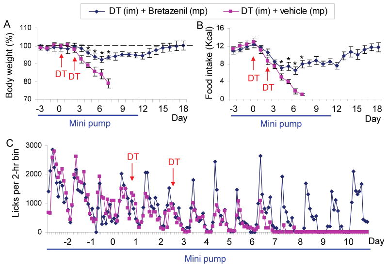
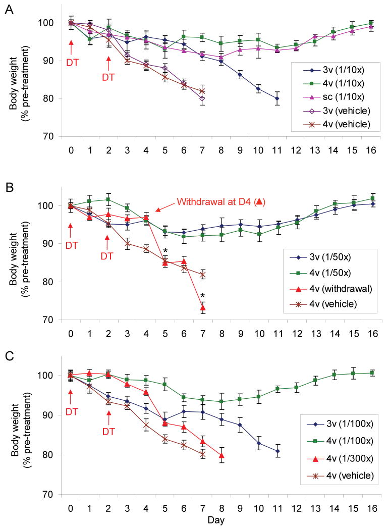
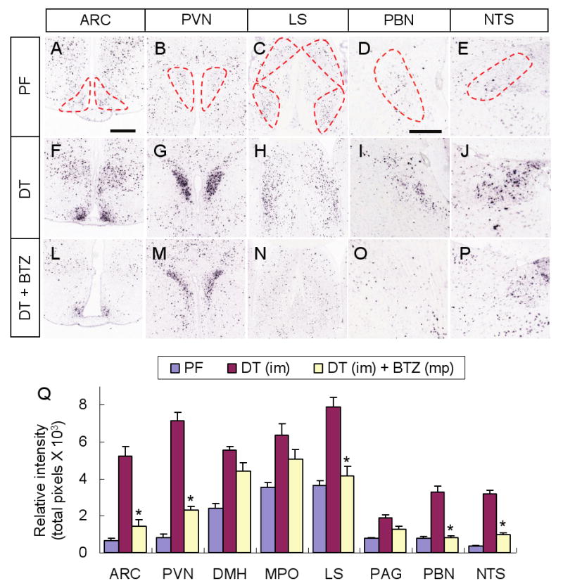
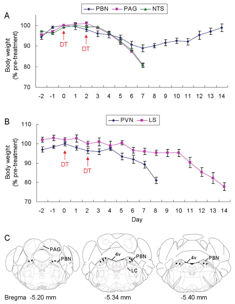
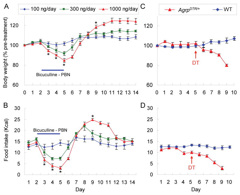
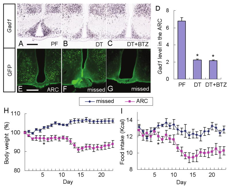
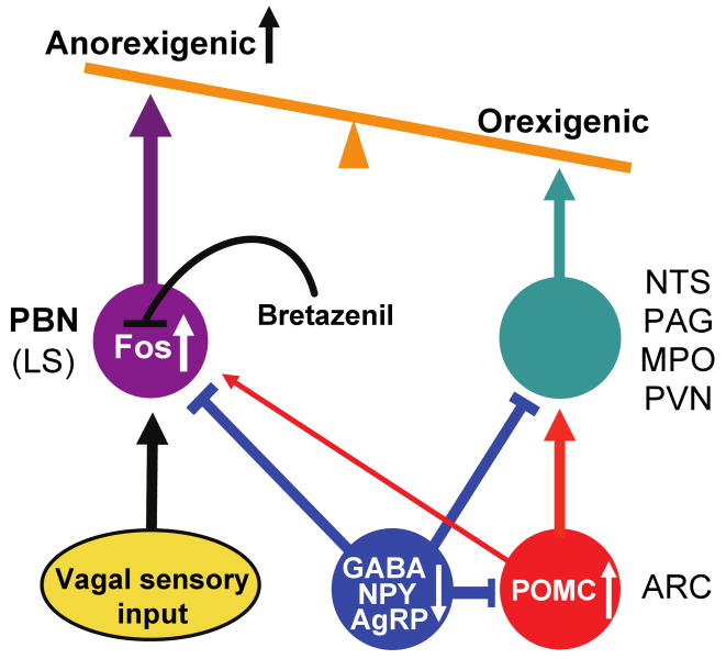
Neurons in the arcuate nucleus that produce AgRP, NPY, and GABA (AgRP neurons) promote feeding. Ablation of AgRP neurons in adult mice results in Fos activation in postsynaptic neurons and starvation. Loss of GABA is implicated in starvation because chronic subcutaneous delivery of bretazenil (a GABA(A) receptor partial agonist) suppresses Fos activation and maintains feeding during ablation of AgRP neurons. Moreover, under these conditions, direct delivery of bretazenil into the parabrachial nucleus (PBN), a direct target of AgRP neurons that also relays gustatory and visceral sensory information, is sufficient to maintain feeding. Conversely, inactivation of GABA biosynthesis in the ARC or blockade of GABA(A) receptors in the PBN of mice promote anorexia. We suggest that activation of the PBN by AgRP neuron ablation or gastrointestinal malaise inhibits feeding. Chronic delivery of bretazenil during loss of AgRP neurons provides time to establish compensatory mechanisms that eventually allow mice to eat.


Figures

Figure 1
Figure 1. Chronic administration of bretazenil protects against starvation in adult mice after acute ablation of AgRP neurons
(A) Percentage of initial body weight of DT-treated, AgrpDTR/+ mice after subcutaneous implantation of mini-osmotic pumps (mp) loaded with either bretazenil (n = 12) or vehicle (n = 10). DT was injected intramuscularly (im) twice as indicated by arrows. Minipumps were removed on Day 11 after drug solution was depleted. (B) Liquid diet intake by the mice described in A. (C) Licking activity of the mice described in A was plotted as average number of licks in 2-h bins. *, p < 0.01, ANOVA. Error bars represent the standard error of mean (SEM).
Figure 2
Figure 2. Minipump delivery of bretazenil into the 4th ventricle is more efficacious than delivery into the 3rd ventricle
(A) Percentage of initial body weight of DT-treated, AgrpDTR/+ mice in which bretazenil (75 ng/hr; 1/10th of the dosage used in Figure 1) or vehicle was chronically administered either subcutaneously (sc) or directly into the 3rd (3v) or 4th (4v) ventricle. (B) Percentage of initial body weight of DT-treated, AgrpDTR/+ mice in which bretazenil (15 ng/hr; 1/50th of the dosage used in Figure 1) or vehicle was chronically administered directly into the 3rd or 4th ventricle. Note that abrupt withdrawal of bretazenil delivered into the 4th ventricle at Day 4 resulted in more profound decline of body weight than that was shown by the AgRP-ablated mice infused with vehicle; *, p < 0.01, ANOVA. (C) Percentage of initial body weight of DT-treated, AgrpDTR/+ mice in which bretazenil (7.5 or 2.5 ng/hr, 1/100th or 1/300th of the dosage used in Figure 1, respectively) or vehicle was chronically administered directly into either the 3rd or 4th ventricle. N = 6 - 8 for each group. Error bars represent the SEM.
Figure 3
Figure 3. Fos activation after AgRP neuron ablation is suppressed in some brain regions by bretazenil treatment
(A-E) Representative pictures of Fos in situ hybridization in post-synaptic regions of AgRP neurons including the ARC (A), PVN (B), LS (C), PBN (D), and NTS (E), in pair-fed AgrpDTR/+ mice after losing ∼20% of their initial body weight. Post-synaptic areas of AgRP neurons are denoted by dotted lines. (F-J) Representative pictures of Fos in situ hybridization in the same post-synaptic regions of AgRP neurons in DT-treated, AgrpDTR/+ mice after losing ∼20% of their initial body weight. (L-P) Representative pictures of Fos in situ hybridization in the same post-synaptic regions of AgRP neurons in DT-treated, AgrpDTR/+ mice with subcutaneous implantation of minipumps eluting bretazenil. (Q) Quantified results for Fos in situ signals in selected post-synaptic regions of AgRP neurons of either pair-fed, DT-treated, or DT/bretazenil-treated, AgrpDTR/+ mice. See Supplementary Figure S6 for a complete set of images of Fos in situ hybridization in these regions. *, p < 0.01 between the DT-treated group and the DT/bretazenil-treated group for each respective area, ANOVA. N = 4 - 6 per group. Scale bar (in A): A-C, F-H, and L-N, 400 μm; scale bar (in D): D, E, I, J, O, and P, 400 μm. Error bars in Q represent the SEM.
Figure 4
Figure 4. Administration of bretazenil into the PBN prevents starvation in mice after ablation of AgRP neurons
(A) Percentage of initial body weight of DT-treated, AgrpDTR/+ mice after chronic administration of bretazenil in 3 post-synaptic areas of AgRP neurons in the hindbrain (PBN, PAG, and NTS). (B) Percentage of initial body weight of DT-treateted, AgrpDTR/+ mice after chronic administration of bretazenil in 2 forebrain regions (PVN and LS) with projections from AgRP neurons. N = 6 – 8 per each group. Error bars represent the SEM. (C) Coronal illustrations of mouse hindbrain regions; the dots represent effective infusion sites in the PBN area. LC, locus ceruleus; 4v, 4th ventricle.
Figure 5
Figure 5. Inhibition of GABAA receptors in the PBN or disruption of the neural inputs from AgRP neurons to the PBN produces anorexia
(A) Percentage of initial body weight of C57BL/6 wild-type mice in which bicuculline (100, 300, and 1000 ng/day) was chronically infused to the PBN area through bilateral cannulae for 4 days and then discontinued. (B) Intake of liquid diet by the mice described in A. *, p < 0.01, ANOVA between the groups of 100 ng/day and 1000 ng/day. Error bars represent the SEM. N = 5 – 6 mice per group. (C) Percentage of initial body weight of AgrpDTR/+ and wild-type mice that received a single injection of DT into the PBN area (bilateral, 4 ng per side). (D) Intake of liquid diet by the mice described in C.
Figure 6
Figure 6. Loss of GABAergic signaling by AgRP neurons promotes anorexia
(A-C) Representative pictures of Gad1 in situ hybridization in the ARC of AgrpDTR/+ mice after either pair feeding (A), DT treatment (B), or DT treatment plus chronic administration of bretazenil (C). (D) Quantified results for Gad1 in situ signals in the ARC of the mice described in A-C. *, p < 0.01, ANOVA. Error bars represent the SEM. N = 6 mice per each group. Scale bar (in A): A-C, 400 μm. (E-G) Representative pictures of GFP immunostaining showing the AAV1-Cre-GFP infected neurons located either precisely in the ARC (n = 4; E) or at nonspecific surrounding areas (F) or completely absent from region (G) (n = 12) in Gad1lox/lox, Gad2-/-, Rosa26fsLacZ/fsLacZ mice. Scale bar (in E): E-G-, 400 μm. (H, I) Percentage change of body weight and intake of liquid diet by the mice with infection in the ARC (E) or missed (F,G). Statistical significance (*, p < 0.01, ANOVA) was obtained starting from Day 6 until end of the experiment. Error bars represent the SEM.
Figure 7
Figure 7. Diagram illustrating how loss of GABAergic signaling from AgRP neurons leads to starvation
AgRP and POMC neurons that reside in the ARC send axons to many of the same target areas, e.g. the PVN, MPO, PAG, NTS and PBN. Ablation (or inhibition) of inhibitory AgRP neurons leads to anorexia by two mechanisms. First, the loss of inhibition onto POMC neurons and their post-synaptic target cells stimulates the melanocortin system, which suppresses feeding. Second, the loss of GABAergic inhibition onto neurons in the PBN (and to a lesser extent the LS) mimics the activation of these neurons in response to gastrointestinal malaise, resulting in severe anorexia, an effect that is independent of melanocortin signaling. The severe anorexia can be prevented by infusing bretazenil, a GABAA receptor partial agonist, into the PBN which antagonizes Fos activation.

Comment in

Similar articles

Cited by

References

    1. Andre V, Dube C, Francois J, Leroy C, Rigoulot MA, Roch C, Namer IJ, Nehlig A. Pathogenesis and pharmacology of epilepsy in the lithium-pilocarpine model. Epilepsia. 2007;48 5:41–47. - PubMed
    1. Bernardis LL, Bellinger LL. The lateral hypothalamic area revisited: ingestive behavior. Neurosci Biobehav Rev. 1996;20:189–287. - PubMed
    1. Berridge KC, Pecina S. Benzodiazepines, appetite, and taste palatability. Neurosci Biobehav Rev. 1995;19:121–131. - PubMed
    1. Broberger C, Hokfelt T. Hypothalamic and vagal neuropeptide circuitries regulating food intake. Physiol Behav. 2001;74:669–682. - PubMed
    1. Broberger C, Johansen J, Johansson C, Schalling M, Hokfelt T. The neuropeptide Y/agouti gene-related protein (AGRP) brain circuitry in normal, anorectic, and monosodium glutamate-treated mice. Proc Natl Acad Sci U S A. 1998;95:15043–15048. - PMC - PubMed

Publication types

Substances