. 2009 Jun 26;137(7):1293-307.
doi: 10.1016/j.cell.2009.04.025.

Mechanism for activation of the EGF receptor catalytic domain by the juxtamembrane segment

Affiliations

Mechanism for activation of the EGF receptor catalytic domain by the juxtamembrane segment

Natalia Jura et al. Cell. .

Erratum in

  • Cell. 2009 Aug 7;138(3):604

Abstract

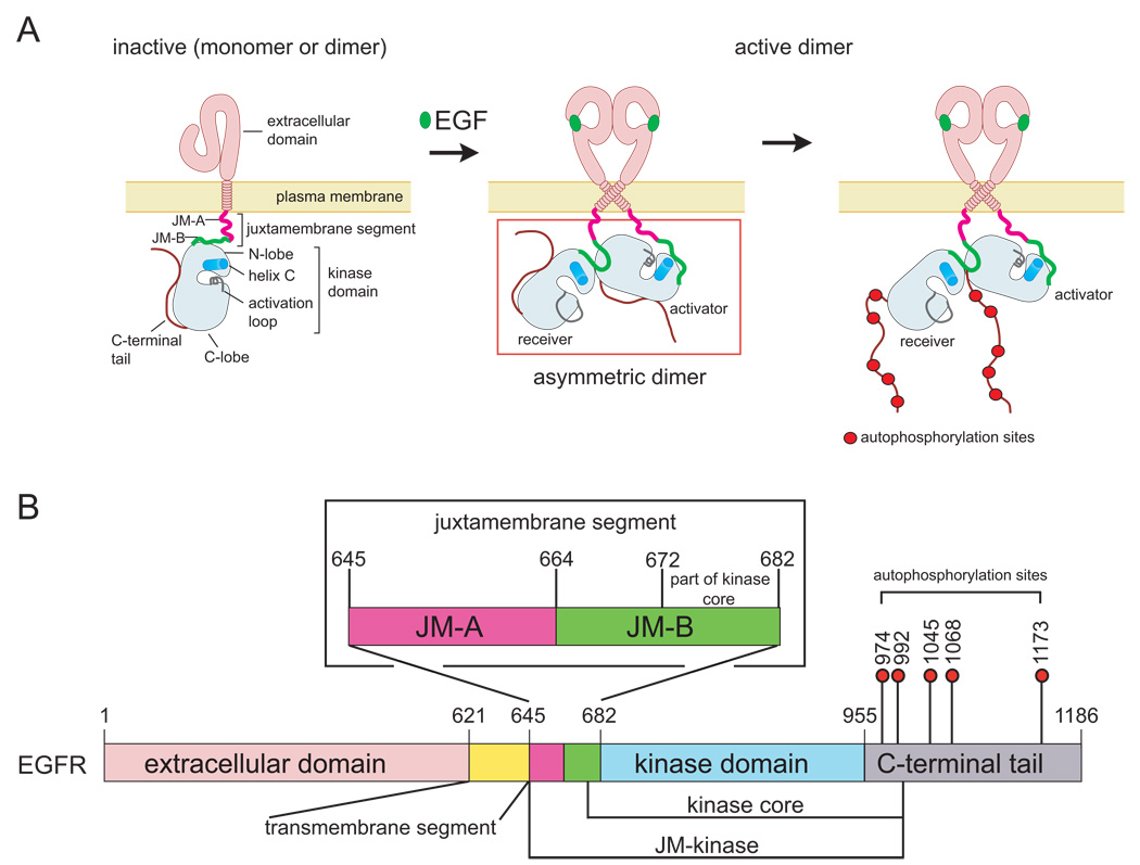
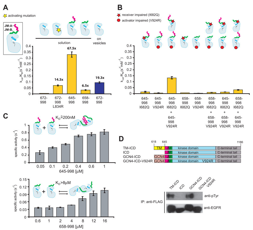
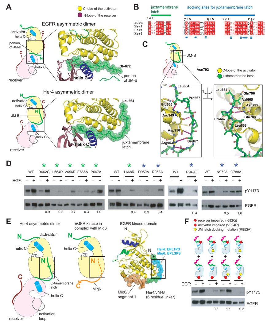
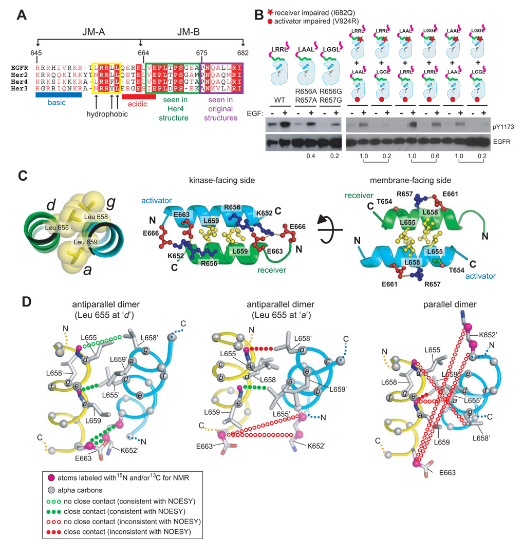
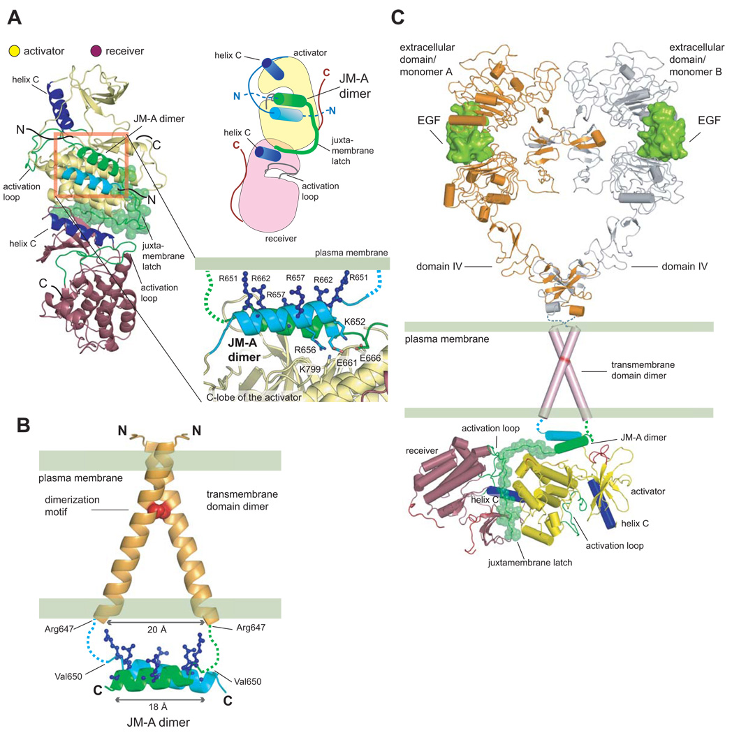
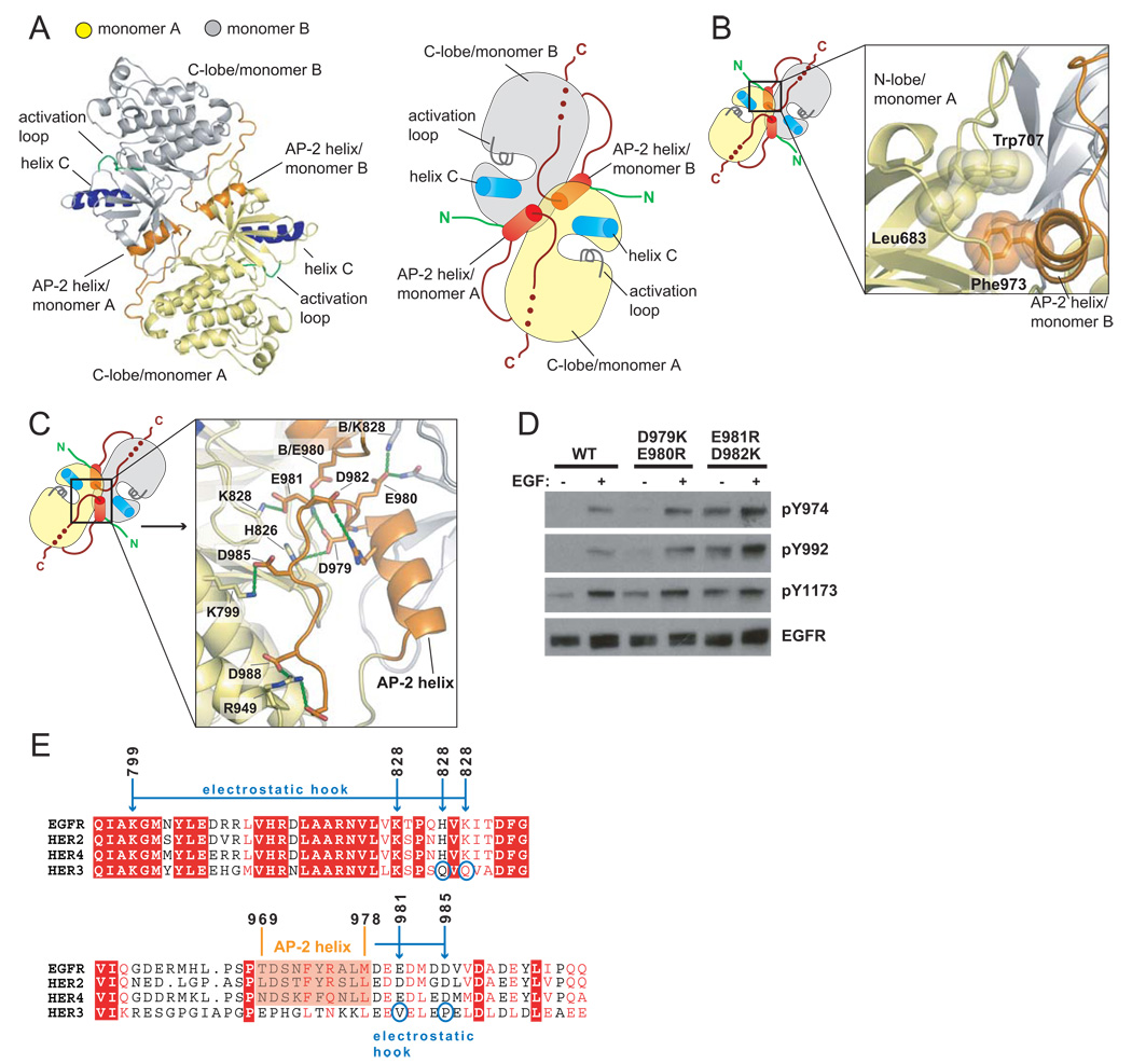
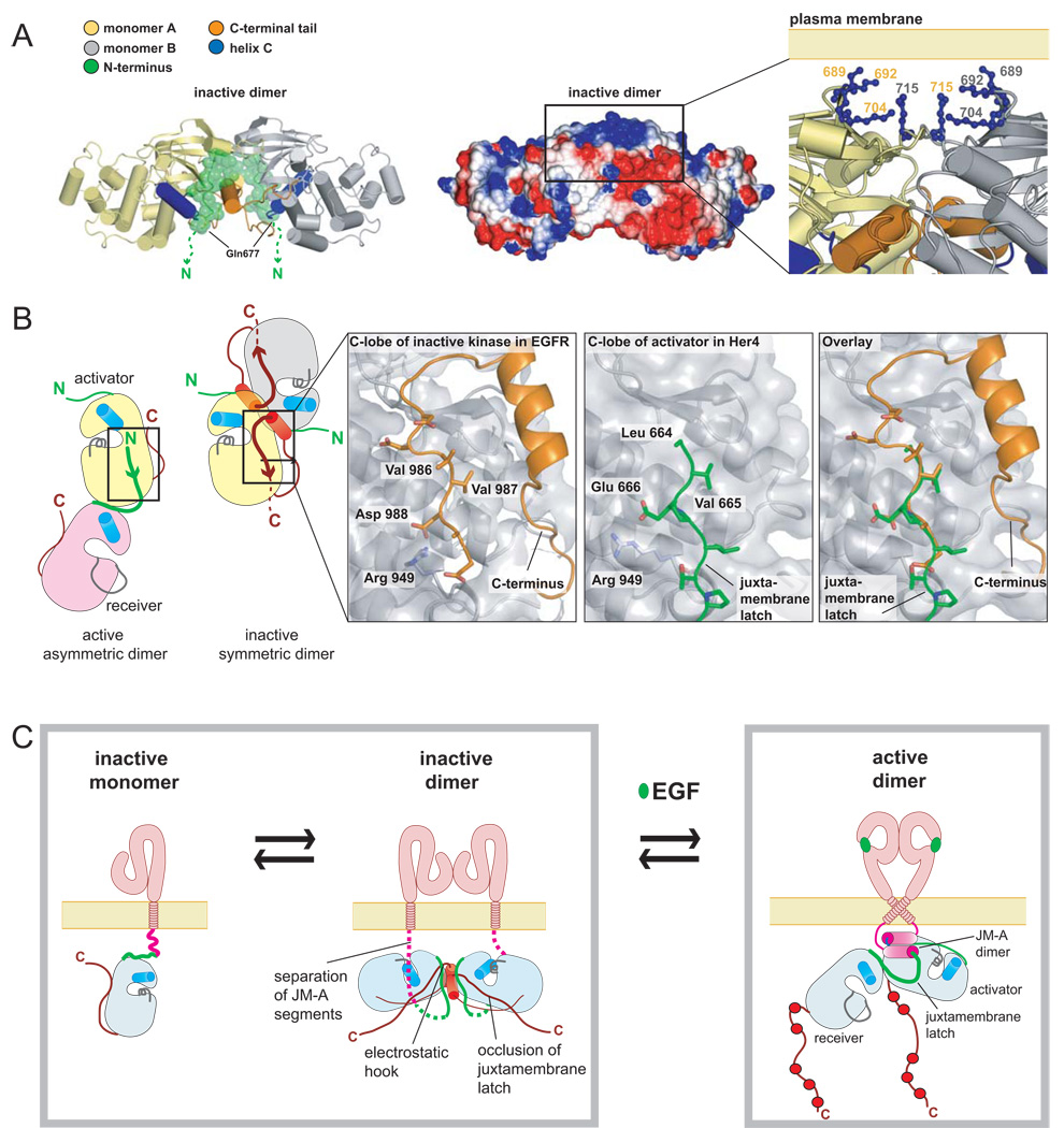
Signaling by the epidermal growth factor receptor requires an allosteric interaction between the kinase domains of two receptors, whereby one activates the other. We show that the intracellular juxtamembrane segment of the receptor, known to potentiate kinase activity, is able to dimerize the kinase domains. The C-terminal half of the juxtamembrane segment latches the activated kinase domain to the activator, and the N-terminal half of this segment further potentiates dimerization, most likely by forming an antiparallel helical dimer that engages the transmembrane helices of the activated receptor. Our data are consistent with a mechanism in which the extracellular domains block the intrinsic ability of the transmembrane and cytoplasmic domains to dimerize and activate, with ligand binding releasing this block. The formation of the activating juxtamembrane latch is prevented by the C-terminal tails in a structure of an inactive kinase domain dimer, suggesting how alternative dimers can prevent ligand-independent activation.


Figures

Figure 1
Figure 1. Schematic Diagrams of EGFR
(A) Activation of EGFR by EGF results in the formation of an asymmetric kinase domain dimer. (B) Domains of EGFR. Residue numbering corresponds to human EGFR, excluding the signal sequence.
Figure 2
Figure 2. The Effect of the Juxtamembrane Segment on Activity
(A) Catalytic efficiency (kcat/KM) of the kinase core (residues 672–998) in solution (yellow) and on vesicles (blue), compared to the catalytic efficiency in solution of constructs that include the full juxtamembrane segment (JM-A and JM-B, residues 645–998) or only JM-B (residues 658–998). The values of kcat/KM were obtained from the linear dependence of reaction velocity on substrate concentration at low substrate concentration, and the error bars are derived from the linear fit (Zhang et al., 2006). (B) The activity of constructs that are either receiver-impaired (restricted to serve as activators, with the I682Q mutation) or activator-impaired (restricted to serve as receivers, with the V924R mutation). (C) Concentration-dependent change in specific activity, in solution, for the JM-kinase construct (containing both JM-A and JM-B, residues 645–998) and a construct containing JM-B but lacking JM-A (residues 658–998). Data shown are mean values from two independent experiments ± STD. (D) EGFR constructs were immunoprecipitated from cell lysates using an anti-FLAG antibody and EGFR autophosphorylation was examined by immunobloting using an anti-phosphotyrosine antibody (anti-pTyr).
Figure 3
Figure 3. Role of the Juxtamembrane Latch in Activation of EGFR
(A) Comparison of the structures of asymmetric dimers of kinase domains for EGFR (PDB ID: 2GS6), (Zhang et al., 2006) and Her4 (PDB ID: 2R4B), (Wood et al., 2008). Residues are identified using EGFR numbering. (B) Sequence conservation in the juxtamembrane latch/C-lobe interface. Residues interacting with the juxtamembrane latch are indicated by asterisks. (C) Detailed view of the structure of the juxtamembrane latch in the Her4 structure (PDB ID: 2R4B), with residues identified by EGFR numbering. (D) Effect of mutating residues involved in formation of the juxtamembrane latch. The level of EGF-stimulated phosphorylation on Tyr 1173 relative to wild type, after normalizing for EGFR levels, is shown below each lane. (E) Comparison of the juxtamembrane latch with the docking of the EGFR inhibitor, Mig6 (PDB ID: 2RFE). (F) The effect of a mutation that prevents docking of the juxtamembrane latch (R953A). The results of co-transfection experiments using full length EGFR receptor variants that are receiver-impaired (I682Q) or activator-impaired (V924R) are shown. The level of EGF-stimulated phosphorylation relative to I682Q and V924R co-transfection in the wild type background, after normalizing for EGFR levels, is shown below each lane.
Figure 4
Figure 4. A Helical Dimer in the JM-A Segment
(A) Alignment of the sequences of the juxtamembrane segments of EGFR family members. (B) Comparison of the effects of alanine and glycine substitutions in the JM-A segment. The first three panels compare activity of the full length wild type receptor with that for variants in which Arg 656 and Arg 657 are replaced by alanine and glycine. The next six panels shows results of co-transfection experiments using activator-impaired and receiver-impaired variants of the receptor, and compare the results of alanine and glycine substitutions in each variant. (C) The modeled antiparallel JM-A helical dimer, with Leu655 at the d position of a heptad motif in one helix and Leu658 and Leu659 at the g and a positions in the second helix. (D) Models for antiparallel (left and middle, with Leu 655 at the d and a positions, respectively) and parallel (right, with Leu 655 at the d position). The dotted lines indicate interatomic contacts that are either consistent or inconsistent with NMR data for a peptide containing two tandem repeats of the JM-A segment (see Supplemental Data).
Figure 5
Figure 5. Structural Coupling Between the Extracellular and Intracellular Domains in Active EGFR
(A) A model for the JM-A helical dimer in the context of the asymmetric dimer of kinase domains. In the exploded view, arginine sidechains that face the membrane are shown. (B) Structure of the transmembrane domain dimer of Her2 (PDB ID: 2JWA), (Bocharov et al., 2008) and the modeled JM-A dimer. Positively charged sidechains that face the membrane are in blue. (C) A model for the activated EGF receptor. Two liganded EGFR extracellular domains are shown in an active dimeric assembly (PDB ID: 1IVO) (Ogiso et al., 2002), with domains IV based on the structure of the inactive EGFR extracellular domain (PDB ID: 1NQL) (Ferguson et al., 2003). This arrangement is compatible with the transmembrane domain dimer and couples to the asymmetric kinase domain dimer via the dimeric JM-A helices and the juxtamembrane latch.
Figure 6
Figure 6. A Symmetric Inactive Dimer of the EGFR Kinase Domain
(A) Overview of the crystal structure of the symmetric inactive dimer. (B) Detailed view of the hydrophobic packing between the C-terminal AP-2 helix of monomer B and the N-lobe of monomer A. (C) Exploded view of the electrostatic hook formed between the C-terminal tail (residues 979–990) of EGFR and the hinge region in the kinase domain. (D) Effect of mutations in the electrostatic hook on autophosphorylation of full length EGFR in COS7 cells. (E) Alignment of the sequences of EGFR family members in the C-terminal tail regions encompassing residues in the electrostatic hook and AP-2 helix.
Figure 7
Figure 7. Proposed Role of the Inactive Dimer in EGFR Autoinhibition
(A) The surface electrostatic potential of the inactive dimer, with positively and negatively charged regions in blue and red, respectively. The exploded view shows the proposed docking at the plasma membrane. (B) Schematic diagram comparing the juxtamembrane latch in the active asymmetric dimer and the docking of the C-terminal tail in the inactive symmetric dimer. (C) A schematic representation of the activation mechanism of EGFR.

Comment in

References

    1. Adams PD, Grosse-Kunstleve RW, Hung LW, Ioerger TR, McCoy AJ, Moriarty NW, Read RJ, Sacchettini JC, Sauter NK, Terwilliger TC. PHENIX: building new software for automated crystallographic structure determination. Acta Crystallogr D Biol Crystallogr. 2002;58:1948–1954. - PubMed
    1. Bell CA, Tynan JA, Hart KC, Meyer AN, Robertson SC, Donoghue DJ. Rotational coupling of the transmembrane and kinase domains of the Neu receptor tyrosine kinase. Mol Biol Cell. 2000;11:3589–3599. - PMC - PubMed
    1. Bocharov EV, Mineev KS, Volynsky PE, Ermolyuk YS, Tkach EN, Sobol AG, Chupin VV, Kirpichnikov MP, Efremov RG, Arseniev AS. Spatial structure of the dimeric transmembrane domain of the growth factor receptor ErbB2 presumably corresponding to the receptor active state. J Biol Chem. 2008;283:6950–6956. - PubMed
    1. Burgess AW, Cho HS, Eigenbrot C, Ferguson KM, Garrett TP, Leahy DJ, Lemmon MA, Sliwkowski MX, Ward CW, Yokoyama S. An open-and-shut case? Recent insights into the activation of EGF/ErbB receptors. Mol Cell. 2003;12:541–552. - PubMed
    1. Burke CL, Lemmon MA, Coren BA, Engelman DM, Stern DF. Dimerization of the p185neu transmembrane domain is necessary but not sufficient for transformation. Oncogene. 1997;14:687–696. - PubMed

Publication types

Associated data