. 1991 Nov 28;354(6351):308-11.
doi: 10.1038/354308a0.

Peripheral deletion of self-reactive B cells

Affiliations

Peripheral deletion of self-reactive B cells

D M Russell et al. Nature. .

Abstract

B LYMPHOCYTES are key participants in the immune response because of their specificity, their ability to take up and present antigens to T cells, and their capacity to differentiate into antibody-secreting cells. To limit reactivity to self antigens, autospecific B cells can be functionally inactivated or deleted. Developing B cells that react with membrane antigens expressed in the bone marrow are deleted from the peripheral lymphocyte pool. It is important to ascertain the fate of B cells that recognize membrane autoantigens expressed exclusively on peripheral tissues because B cells in the peripheral lymphoid organs are phenotypically and functionally distinct from bone-marrow B cells. Here we show that in immunoglobulin-transgenic mice, B cells specific for major histocompatibility complex class I antigen can be deleted if they encounter membrane-bound antigen at a post-bone-marrow stage of development. This deletion may be necessary to prevent organ-specific autoimmunity.


Figures

FIG. 1
FIG. 1
Maps of the DNA fragments used to generate 3-83μδ transgenic mice. Dark regions represent exons. METHODS. Mice were produced using functional rearranged heavy- and light-chain genes encoding the 3-83 antibody. This antibody binds with moderate affinity to Kk, with weak affinity to Kb, and fails to bind to H–2d cells. Transgenic mice encoding the IgM form of 3-83 have been described-. Self tolerance to Kk and Kb in the IgM-only 3-83 mice is mediated by deletion in H–2d transgenic → H–2d × H–2k or H–2d transgenic → H–2d × H–2b bone marrow chimaeras,. To generate the 3-83 IgM plus IgD heavy-chain fragment, the 3-83μ construct used previously was extended to include the complete genomic locus. The 3-83μ insert was liberated from its vector by partial digestion with EcoR1 and cloned into the EcoR1 site of λEMBL3 to generate λ241. A cosmid clone spanning the (DBA/2-derived) Ig-Cμ and Ig-Cδ regions was then isolated from a genomic library of the T-cell hybrid BDF1 16 (ref. 23), linearized with XhoI, which cuts at the site between and , and ligated together with the VDJ/Cμ-containing SalI/XhoI fragment of λ 241 and with SalI-digested pNNL cosmid vector, resulting in a cosmid whose restriction map in the Cμ/Cδ region corresponds to the natural locus. The 42-kilobase (kb) insert was liberated from all but ~200 base pairs (bp) of the vector by NruI digestion. Light- and heavy-chain gene fragments were isolated and transgenic mice produced as described. Southern blotting and segregation analysis indicated that the 3-83μδ line has ~3–5 copies of the light- and heavy-chain genes co-integrated at a single chromosomal locus. m, Transmembrane exons.
FIG. 2
FIG. 2
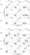
Elimination of autoreactive B cells in spleen and lymph nodes, but not in the bone marrow, of an Ig-Tg mouse bearing peripherally expressed antigen. Cells from the organs of 5-week-old transgenic and normal littermates were analysed by two-colour flow cytometry. a, Staining with anti-idiotype and anti-IgM. Idiotype was detected with the 3-83 clonotype-specific rat monoclonal antibody 54.1, which recognizes an epitope created by the pairing of 3-83 heavy and light chains. b, Staining with anti-IgD and anti-IgM. Dbl-Tg, double transgenic (3-83μγ/MT-Kb); Ig-Tg, immunoglobulin transgenic; Non-Tg, non-transgenic. Cells were prepared as before and stained with monoclonal antibodies: anti-3-83 clonotype, 54.1/biotin; anti-IgM, DS1/FITC (ref. 25); anti-IgDa, AMS 9.1/biotin (Pharmingen). Biotin-conjugated antibodies were revealed with phycoerythrin (PE)–streptavidin stain (Becton–Dickinson). Purification and fluorochrome conjugation of the antibodies was as described. Flow cytometry analysis was on a Profile (Coulter) machine and data are presented using its density plot function. The percentages of cells in each quadrant, rounded to the nearest 0.1%, are indicated in the upper right corner of each plot. Data for bone marrow cells are gated by side scatter to exclude the predominant myeloid cell population from analysis. All mice used in this experiment were H–2d and Igh-Ca at the endogenous loci. The genetic background of the mice is ~75% BALB/cJ, 6% B10.D2, 6% C57b16/J, 6% SJL/J, 6% DBA/2. The transgenic havy chain μ and δ allotypes are type a. The results shown are representative of 4 experiments (Table 1). MT-Kb mice were indistinguishable from Non-Tg mice in these assays (not shown).
FIG. 3
FIG. 3
RNA analysis of transgenic and non-transgenic mice. a, Northern blot analysis shows reduction in transgenic κ-light chain expression in spleen and lymph nodes of Dbl-Tg mice. After hybridization with Vκ 3-83 probe and decay of the radioactive signal, the filter was hybridized with the MHC class 1-specific pll2a probe. b, Polymerase chain reaction (PCR) detection of Vκ 3-83 RNA in the livers of peripherally deleting mice. Lanes 1, 3, 5 and 7, liver; lanes 2, 4, 6 and 8, bone marrow. Lanes 1 and 2, mouse 2080 (Dbl-Tg); lanes 3 and 4, mouse 2081 (Dbl-Tg); lanes 5 and 6, mouse 2082 (Ig–Tg); lanes 7 and 8, Non-Tg littermate. Complementary DNA was synthesized and amplified with 15 cycles of PCR, run on a 1.2% agarose gel, transferred to a Zetaprobe membrane (BioRad) and hybridized with probes recognizing gene transcripts of 3-83 Vκ or the GTP-binding protein Gαs (ref. 29). Lane M, marker DNA fragments of 587, 434 and 267 bp. METHODS. RNA preparation and northern blotting were as described. 5μg RNA was loaded per lane in the 1% agarose formaldehyde gel. RNA samples were reverse-transcribed using a commercial kit (Superscript, BRL). PCR reactions were in a final volume of 30 μl and contained 1 μl reverse transcription reaction, 10 mM Tris–HCl, pH 8.3, 50 mM KCl, 1.5 mM MgCl2, 20 μg ml−1 gelatin, 0.3 mM each of dATP, dCTP, dGTP and dTTP, 1 μM of each oligonucleotide primer, and 1 U of Taq polymerase (Perkin–Elmer Cetus). Each cycle consisted of 30 s at 94 °C, 30 s at 62 °C and 1.5 min at 72 °C in a thermal cycler (Ericomp). The appropriate cDNA clones were used as hybridization probes after labelling with [α-32P)dCTP. PCR primers were: 5′Vκ 3-83 (79-101): 5′-CAGCTTCCTGCTAATCAGTGCC-3′; 3′Jκ2 (412-431): 5′-TGGTCCCCCCTCCGAACGTG-3′; Gαs sense (309-336): 5′-ATTGAAACCATTGTGGCCGCCATGAGC-3′; Gαs antisense (1,104-1,128): 5′-GAAGACACGGCGGATGTTCTCAGTGTC-3′.

References

    1. Nossal GJV. Science. 1989;24:147–153. - PubMed
    1. Goodnow CC, et al. Nature. 1988;334:676–682. - PubMed
    1. Erikson J, et al. Nature. 1991;349:331–334. - PubMed
    1. Nemazee D, Bürki K. Nature. 1989;337:562–566. - PubMed
    1. Nemazee D, Bürki K. Proc natn Acad Sci U S A. 1989;86:8039–8043. - PMC - PubMed

Publication types