Review
. 2009 Jul;5(3):349-67, vi.
doi: 10.1016/j.hfc.2009.02.009.

Identifying the etiology: a systematic approach using delayed-enhancement cardiovascular magnetic resonance

Affiliations
Review

Identifying the etiology: a systematic approach using delayed-enhancement cardiovascular magnetic resonance

Annamalai Senthilkumar et al. Heart Fail Clin. 2009 Jul.

Abstract

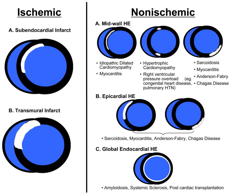
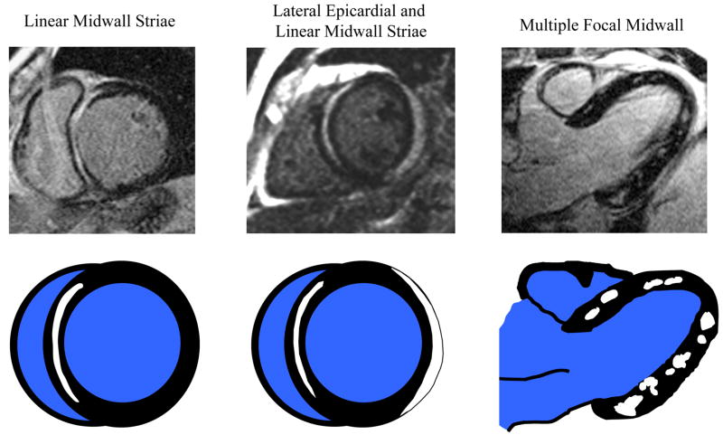
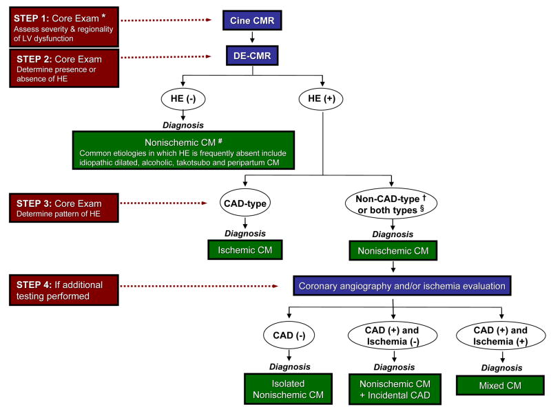
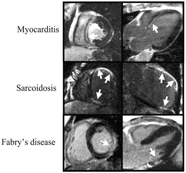
In patients who have heart failure, treatment and survival are directly related to the cause. Clinically, as a practical first step, patients are classified as having either ischemic or non-ischemic cardiomyopathy, a delineation usually based on the presence or absence of epicardial coronary artery disease. However, this approach does not account for patients with non-ischemic cardiomyopathy who also have coronary artery disease, which may be either incidental or partly contributing to myocardial dysfunction (mixed cardiomyopathy). By allowing direct assessment of the myocardium, delayed-enhancement cardiovascular magnetic resonance (DE-CMR) may aid in addressing these conundrums. This article explores the use of DE-CMR in identifying ischemic and non-ischemic myopathic processes and details a systematic approach to determine the cause of cardiomyopathy.


Figures

Figure 1
Figure 1
Unadjusted Kaplan-Meier survival curves for ischemic versus nonischemic cardiomyopathy. Reproduced with permission from Felker GM, Shaw LK, O’Connor CM. A standardized definition of ischemic cardiomyopathy for use in clinical research. J Am Coll Cardiol. 2002;39(2):210–218.
Figure 2
Figure 2
Images before and after coronary stenting demonstrate the ability of delayed enhancement cardiac magnetic resonance to identify micro-infarcts. Arrows point to new discrete regions of hyperenhancement in the inferior wall related to the procedure. Adapted with permission from Ricciardi MJ, Wu E, Davidson CJ, Choi KM, Klocke FJ, Bonow RO, Judd RM, Kim RJ. Visualization of discrete microinfarction after percutaneous coronary intervention associated with mild creatine kinase-MB elevation. Circulation. 2001;103(23):2780–2783.
Figure 3
Figure 3
Mechanisms of hyperenhancement in myocardial infarction: Scar may be the result of chronic infarction or nonischemic damage (see text for details). Adapted from Kim RJ, Elliott MD, Judd RM. Assessment of Myocardial Viability by Contrast Enhancement. In: Higgins CB, de Roos A, eds. MRI and CT of the Cardiovascular System. 2nd ed. Philadelphia, PA: Lippincott Williams & Wilkins; 2005.
Figure 4
Figure 4
The typical hyperenhancement pattern of myocardial infarction can be explained by the pathophysiology of ischemia. Little or no cellular necrosis is found until about 15 min after occlusion. Over the next few hours a wavefront of necrosis begins in the subendocardium and moves progressively towards the epicardium. During this period, the infarcted region within the ischemic zone increases continuously and can ultimately become transmural. Reproduced with permission from Mahrholdt H, Wagner A, Judd RM, Sechtem U, Kim RJ. Delayed enhancement cardiovascular magnetic resonance assessment of non-ischaemic cardiomyopathies. Eur Heart J. 2005;26(15):1461–1474.
Figure 5
Figure 5
Hyperenhancement patterns that one may encounter in clinical practice. If hyperenhancement is present, the endocardium should be involved in patients with ischemic disease. Isolated midwall or epicardial hyperenhancement strongly suggests a nonischemic etiology. Reproduced with permission from Shah DJ, Judd RM, Kim RJ. Myocardial Viability. In: Edelman RR, Hesselink JR, Zlatkin MB, Crues JV, eds. Clinical Magnetic Resonance Imaging. 3rd ed. New York, NY: Elsevier; 2006.
Figure 6
Figure 6
The top row displays delayed enhancement images of three commonly encountered patterns of hyperenhancement in patients with myocarditis. The bottom row is a schematic representation of the respective hyperenhancement patterns.
Figure 7
Figure 7
A proposed stepwise approach for evaluating patients with heart failure incorporating delayed enhancement cardiovascular magnetic resonance. CAD=coronary artery disease; CM=cardiomyopathy; CMR=cardiac magnetic resonance; DE=delayed enhancement; HE=hyperenhancement; LV=left ventricle. * core exam typically consists of cine followed by delayed-enhancement imaging. # presumes long-standing, chronic cardiomyopathy and severe ventricular dysfunction. † see Table 1 and Figure 5 for details of non-CAD-type patterns of HE in various nonischemic cardiomyopathies. § often patients with nonischemic CM can have both non-CAD and CAD type HE (e.g. sarcoidosis)
Figure 8
Figure 8
Hyperenhancement (arrows) in nonischemic cardiomyopathies can sometimes mimic that of coronary artery disease. The patient in the upper row had biopsy proven HHV6 myocarditis and no coronary artery disease by angiography. Cardiac sarcoidosis can cause transmural scarring resulting in wall thinning just as myocardial infarction (middle row). The lesions of Anderson-Fabry disease are usually intramural, except for few cases where also the subendocardium can be affected (bottom row). Bottom row, modified with permission from Moon JC, Sachdev B, Elkington AG, McKenna WJ, Mehta A, Pennell DJ, Leed PJ, Elliott PM. Gadolinium enhanced cardiovascular magnetic resonance in Anderson-Fabry disease. Evidence for a disease specific abnormality of the myocardial interstitium. Eur Heart J. 2003;24(23):2151–2155.
Figure 9
Figure 9
The most basal aspect of a myocardial infarction can spare the subendocardium as illustrated by gross pathology in an canine heart with acute MI (top row; modified with permission from Kim RJ, Fieno DS, Parrish TB, Harris K, Chen EL, Simonetti O, Bundy J, Finn JP, Klocke FJ, Judd RM. Relationship of MRI delayed contrast enhancement to irreversible injury, infarct age, and contractile function. Circulation. 1999;100(19):1992–2002) and in vivo delayed enhancement images from a patient with acute infarction (bottom row); arrows point to infarcted myocardium. Examining the more distal contiguous slices and orthogonal views demonstrates the subendocardial involvement by the infarction. Thus, before deciding that hyperenhancement is non-CAD-type, e.g. “spares the subendocardium”, note that subendocardial sparing refers to the entire extent of hyperenhancement and not merely a small portion.
Figure 10
Figure 10
It may be difficult to differentiate normal myocardium from no-reflow zones within the core of an acute MI (arrow, Panel A) using inversion times to null the normal myocardium since both regions appear black. However, with higher inversion times (~600 ms), the no-reflow region which remains black, may be reliably differentiated from normal myocardium, which is now gray in appearance (Panel B).

References

    1. Richardson P, McKenna W, Bristow M, Maisch B, Mautner B, O’Connell J, Olsen E, Thiene G, Goodwin J, Gyarfas I, Martin I, Nordet P. Report of the 1995 World Health Organization/International Society and Federation of Cardiology Task Force on the Definition and Classification of cardiomyopathies. Circulation. 1996;93(5):841–842. - PubMed
    1. Maron BJ, Towbin JA, Thiene G, Antzelevitch C, Corrado D, Arnett D, Moss AJ, Seidman CE, Young JB. Contemporary definitions and classification of the cardiomyopathies: an American Heart Association Scientific Statement from the Council on Clinical Cardiology, Heart Failure and Transplantation Committee; Quality of Care and Outcomes Research and Functional Genomics and Translational Biology Interdisciplinary Working Groups; and Council on Epidemiology and Prevention. Circulation. 2006;113(14):1807–1816. - PubMed
    1. Alderman EL, Fisher LD, Litwin P, Kaiser GC, Myers WO, Maynard C, Levine F, Schloss M. Results of coronary artery surgery in patients with poor left ventricular function (CASS) Circulation. 1983;68(4):785–795. - PubMed
    1. Felker GM, Shaw LK, O’Connor CM. A standardized definition of ischemic cardiomyopathy for use in clinical research. J Am Coll Cardiol. 2002;39(2):210–218. - PubMed
    1. Packer M, Coats AJ, Fowler MB, Katus HA, Krum H, Mohacsi P, Rouleau JL, Tendera M, Castaigne A, Roecker EB, Schultz MK, DeMets DL. Effect of carvedilol on survival in severe chronic heart failure. N Engl J Med. 2001;344(22):1651–1658. - PubMed

Publication types

MeSH terms