. 2009 Jul 6;206(7):1615-31.
doi: 10.1084/jem.20090490. Epub 2009 Jun 29.

A mutation in Irak2c identifies IRAK-2 as a central component of the TLR regulatory network of wild-derived mice

Affiliations

A mutation in Irak2c identifies IRAK-2 as a central component of the TLR regulatory network of wild-derived mice

James R Conner et al. J Exp Med. .

Abstract

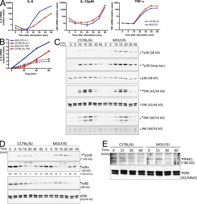
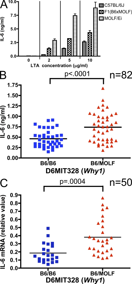
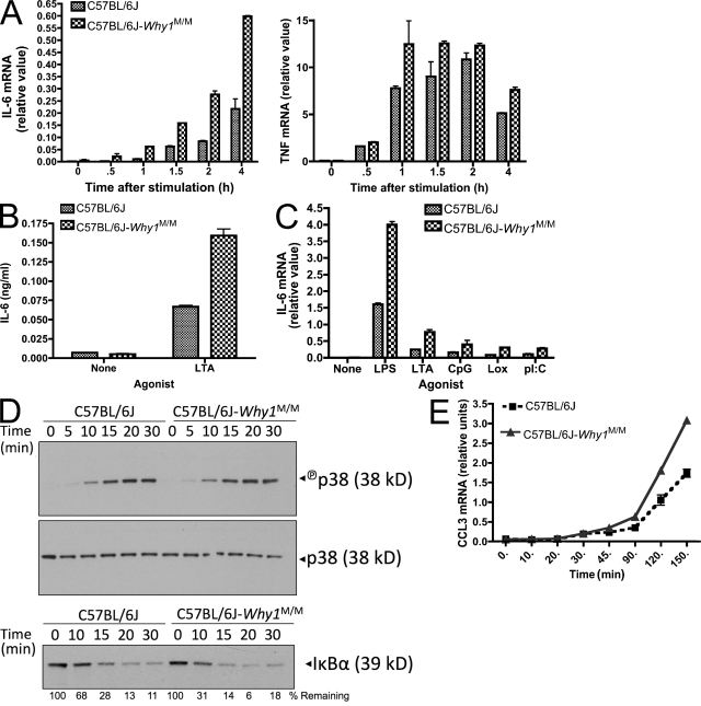
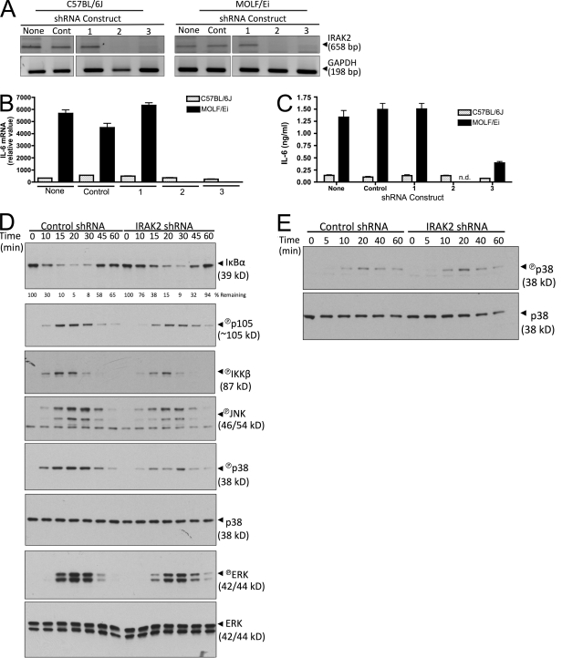
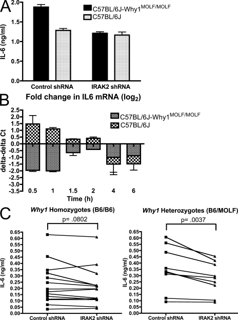
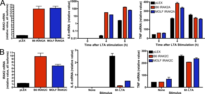
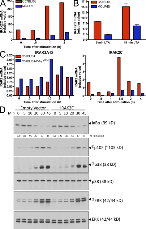
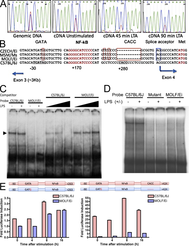
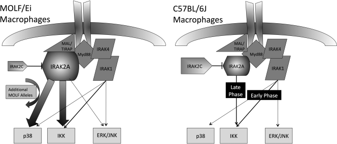
In a phenotypic screen of the wild-derived mouse strain MOLF/Ei, we describe an earlier and more potent toll-like receptor (TLR)-mediated induction of IL-6 transcription compared with the classical inbred strain C57BL/6J. The phenotype correlated with increased activity of the IkappaB kinase axis as well as p38, but not extracellular signal-regulated kinase or c-Jun N-terminal kinase, mitogen-activated protein kinase (MAPK) phosphorylation. The trait was mapped to the Why1 locus, which contains Irak2, a gene previously implicated as sustaining the late phase of TLR responses. In the MOLF/Ei TLR signaling network, IRAK-2 promotes early nuclear factor kappaB (NF-kappaB) activity and is essential for the activation of p38 MAPK. We identify a deletion in the MOLF/Ei promoter of the inhibitory Irak2c gene, leading to an increased ratio of pro- to antiinflammatory IRAK-2 isoforms. These findings demonstrate that IRAK-2 is an essential component of the early TLR response in MOLF/Ei mice and show a distinct pathway of p38 and NF-kappaB activation in this model organism. In addition, they demonstrate that studies in evolutionarily divergent model organisms are essential to complete dissection of signal transduction pathways.


Figures

Figure 1.
Figure 1.
Hyperactivation of cytokines and intracellular mediators in TLR2-stimulated MOLF/Ei macrophages. (A) Peritoneal macrophages from C57BL/6J and MOLF/Ei mice were stimulated with 2 µg/ml LTA for the indicated times. Cytokine mRNA was quantified by real-time PCR. Data represent mean ± range from duplicate wells. One of at least three representative experiments is shown. (B) MEFs were prepared from MOLF/Ei and C57BL/6J mice and stimulated with 100 ng/ml human IL-1β or TNF-α for the indicated times. IL-6 mRNA was quantified using real-time PCR. Data are shown as mean ± range from duplicate wells, representative of two independent experiments. (C–E) BMDMs from each strain were stimulated with 2 µg/ml LTA for the indicated times. Cytoplasmic protein lysates were analyzed by Western blot analysis for MAPK (C) and NF-κB (D) pathway activity or IRAK-1 degradation (E). Data are representative of three (C and D) or two (E) independent experiments.
Figure 2.
Figure 2.
TLR2-stimulated IL-6 production is associated with the Why1 locus. (A) Codominant inheritance of IL-6 protein secretion. Peritoneal macrophages from parental strains and F1(C57BL/6JxMOLF/Ei) hybrid mice were plated and stimulated for 6 h with the indicated concentrations of LTA. IL-6 secretion was measured by ELISA. Results are ± SEM from triplicate wells, representative of three independent experiments. (B) Peritoneal macrophages from 82 N2 (C57BL/6Jx(C57BL/6JxMOLF/Ei)) mice were plated and stimulated with 2 µg/ml LTA as in A. Mean IL-6 production from triplicate wells is plotted for each mouse, grouped by genotype at D6MIT328, a marker near the peak of the Why1 locus. (C) Peritoneal macrophages from 50 independent N2 backcross mice were stimulated for 1 h with 2 µg/ml LTA. IL-6 mRNA was quantified by real time PCR. Mean mRNA production from two wells is plotted, grouped by genotype at D6MIT328. P-values were calculated in B and C using a two-tailed Student's t test.
Figure 3.
Figure 3.
Increased IL-6 production in C57BL/6J-Why1MOLF/MOLF congenic mice. (A and B) Peritoneal macrophages were isolated from wild-type and Why1 congenic mice and stimulated with 2 µg/ml LTA. IL-6 or TNF mRNA was analyzed by real-time PCR at the indicated times (A) and IL-6 protein secretion was analyzed by ELISA 6 h after stimulation (B). (C) Cells were stimulated for 2 h with 100 ng/ml LPS, 2 µg/ml LTA, 200 nM CpG, 100 µM Loxoribine, or 25 µg/ml poly(I:C), and IL-6 mRNA was analyzed by real-time PCR. Data shown as mean ± range of duplicate wells (A and C) or ± SEM of triplicate wells (B). (D) Cells from wild-type and Why1 congenic mice were stimulated with 2 µg/ml LTA for the indicated times and protein was extracted for Western blot analysis of p38 activation and IκBα degradation. Total p38 served as a loading control. (E) Peritoneal macrophages from the indicated strains were stimulated with 2 µg/ml LTA for the indicated times and CCL3 mRNA was measured by real-time PCR analysis. Error bars indicate SEM. All experiments are representative of two to three independent trials.
Figure 4.
Figure 4.
IRAK-2 is required for early TLR2 responses in MOLF/Ei but not C57BL/6J macrophages. (A–C) C57BL/6J and MOLF/Ei BMDMs were infected with lentiviral constructs containing either control or one of three IRAK-2–targeting shRNA sequences. (A) To assess knockdown efficiency, mRNA was harvested and cDNA was amplified with IRAK-2 or GAPDH-specific PCR primers. (B and C) Cells were stimulated with 2 µg/ml LTA and IL-6 was quantified at 4 h using RT-PCR, with error bars indicating SEM (B), or at 6 h using ELISA, with error bars indicating SEM (C). n.d., not detected. Data are representative of three independent experiments. (D and E) MOLF/Ei BMDMs were infected with control or IRAK-2–targeting construct number two and stimulated with 2 µg/ml LTA (D) or under hypertonic NaCl conditions (E) for the indicated times. MAPK and NF-κB pathway signaling events were assessed using the indicated Western blot analyses. Total p38 and ERK serve as loading controls. Data are representative of at least two independent experiments.
Figure 5.
Figure 5.
The effect of IRAK-2 knockdown in macrophages is allele specific. (A and B) BMDMs from C57BL/6J and C57BL/6J-Why1MOLF/MOLF mice were transduced with control or an IRAK-2–targeting shRNA construct. Cells were stimulated with 2 µg/ml LTA for 6 h followed by IL-6 ELISA analysis (A) or for the indicated times followed by IL-6 mRNA quantification by real-time PCR (B). Real-time PCR data are shown as the fold change (in Ct values) observed in IRAK-2 shRNA treated relative to control-treated cells. Data shown as mean ± SEM from triplicate wells (A) or ± range from duplicate wells (B), representative of two independent experiments. (C) Peritoneal macrophages from 14 Why1 homozygous or 9 heterozygous N2 backcross mice were plated and infected with control or IRAK-2–targeting shRNA constructs. 3 d after infection, cells were stimulated with 2 µg/ml LTA and IL-6 protein secretion was measured by ELISA. Mean values of three wells for each mouse are plotted. P-values were calculated using a paired Student's t test analysis.
Figure 6.
Figure 6.
Ectopic expression of MOLF/Ei or C57BL/6J IRAK-2 isoforms results in similar effects on TLR2 activity. RAW264.7 macrophages were transduced with a lentiviral construct encoding the MOLF/Ei or C57BL/6J alleles of IRAK-2A (A) or IRAK-2C (B). Cells were stimulated for the indicated times with 2 µg/ml LTA. Total levels of IRAK-2 in unstimulated cells as well as IL-6 and TNF mRNA accumulation were assessed by real-time PCR. Data are depicted as mean ± range of duplicate wells, representative of three independent experiments.
Figure 7.
Figure 7.
IRAK-2C is differentially expressed between MOLF/Ei and C57BL/6J macrophages and inhibits NF-κB activity. (A) BMDMs from MOLF/Ei and C57BL/6J mice were stimulated for the indicated times with 2 µg/ml LTA and IRAK-2C mRNA was assessed using real-time PCR primers with a forward primer located within the unique 5′ untranslated region of IRAK-2C and a reverse primer located within IRAK-2 exon 6. Primer specificity was confirmed by agarose gel electrophoresis and sequencing (not depicted). (B and C) Peritoneal macrophages from the indicated strains were stimulated for the times shown with 2 µg/ml LTA. IRAK-2C mRNA was quantified as in A and total IRAK-2 mRNA was quantified using primers common to all isoforms (C). Error bars indicate SEM. (D) RAW264.7 cells were transduced with a lentiviral construct coding for the MOLF/Ei allele of IRAK-2C. Cells were stimulated for the indicated times with 2 µg/ml LTA, and NF-κB and MAPK pathways were assessed by Western blot analysis. Total p38 and ERK served as loading controls. All data are representative of two to four independent experiments.
Figure 8.
Figure 8.
Identification of cis-acting regulatory elements conferring differential IRAK-2C expression. (A) Genomic DNA or cDNA from F1(C57BL/6JxMOLF/Ei) macrophages was PCR amplified and analyzed by sequence analysis to determine allelic bias in IRAK-2C transcript. (B) Sequence alignment of the genomic region of IRAK-2C from three wild-derived strains (top three lanes) and C657BL/6J (bottom lane). Positions of the elements are given in base pairs with respect to the transcription initiation site (at 0 bp). Red boxes, mutations common for all wild-derived strains; Blue box, splice acceptor site for IRAK-2A isoforms; red text, NF-κB consensus and translation start of the IRAK-2C protein. Consensus sites were identified using MatInspector software (Genomatix Software GmbH). (C) EMSA was performed using nuclear extracts of LPS-activated RAW cells and a double-stranded 32P-labeled oligonucleotide corresponding to the sequence containing the GATA-binding site. Cold competition assay was performed with 5×-, 25×-, or 100×-fold excess of either B6 or MOLF unlabeled oligonucleotide. (D) Nuclear extracts from LPS-activated RAW cells were incubated with radiolabeled probe derived from C57BL/6J sequence or a sequence with an (A/T) substitution in the CACC-binding site (mutant). Alternatively, a MOLF-derived probe from the region corresponding to the 10-bp deletion was used. (E) RAW264.7 cells were stably transfected with constructs containing the indicated IRAK-2C promoter regions from MOLF/Ei or C57BL/6J fused to luciferase. Equivalent levels of transfection and integration were confirmed using real-time PCR analysis of genomic DNA (not depicted). Cells were stimulated with 100 ng/ml LPS for the indicated times and luciferase was quantified as a measure of promoter activity. Values are expressed relative to unstimulated MOLF/Ei promoter constructs and are shown as mean ± SEM of three wells. Data are representative of three independent experiments.
Figure 9.
Figure 9.
Model of IRAK-2 usage in MOLF/Ei compared with C57BL/6J TLR signal transduction. In MOLF/Ei macrophages, knockdown experiments reveal that IRAK-2 plays a central role in activation of p38 and IKK, two pathways which show increased activity relative to C57BL/6J mice. Neither of these phenotypes can be completely recapitulated with the MOLF/Ei allele on a classical inbred background, suggesting that additional MOLF/Ei alleles are important for the complete levels of hyperresponsiveness observed in comparing parental strains. The MOLF/Ei allele of Irak2 expresses less of the IRAK-2C isoform, which may underlie the phenotype by decreasing interference with IRAK-2A activity. In C57BL/6J macrophages, which express an increased amount of IRAK-2C, previous studies have demonstrated that early TLR signaling events are not controlled by IRAK-2, although it is important for late activation of the NF-κB axis.

References

    1. Akira S., Uematsu S., Takeuchi O. 2006. Pathogen recognition and innate immunity.Cell. 124:783–801 - PubMed
    1. Beebe A.M., Mauze S., Schork N.J., Coffman R.L. 1997. Serial backcross mapping of multiple loci associated with resistance to Leishmania major in mice.Immunity. 6:551–557 - PubMed
    1. Beutler B., Jiang Z., Georgel P., Crozat K., Croker B., Rutschmann S., Du X., Hoebe K. 2006. Genetic analysis of host resistance: toll-like receptor signaling and immunity at large.Annu. Rev. Immunol. 24:353–389 - PubMed
    1. Bonizzi G., Karin M. 2004. The two NF-kappaB activation pathways and their role in innate and adaptive immunity.Trends Immunol. 25:280–288 - PubMed
    1. Burns K., Janssens S., Brissoni B., Olivos N., Beyaert R., Tschopp J. 2003. Inhibition of interleukin 1 receptor/Toll-like receptor signaling through the alternatively spliced, short form of MyD88 is due to its failure to recruit IRAK-4.J. Exp. Med. 197:263–268 - PMC - PubMed

Publication types

MeSH terms