. 2009 Sep;53(9):3914-22.
doi: 10.1128/AAC.00657-09. Epub 2009 Jun 29.

Candida albicans and Staphylococcus aureus form polymicrobial biofilms: effects on antimicrobial resistance

Affiliations

Candida albicans and Staphylococcus aureus form polymicrobial biofilms: effects on antimicrobial resistance

Melphine M Harriott et al. Antimicrob Agents Chemother. 2009 Sep.

Abstract

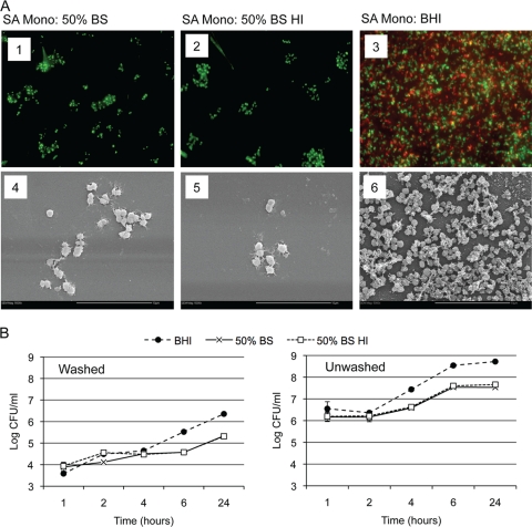
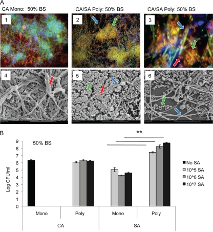
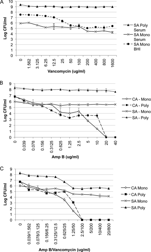
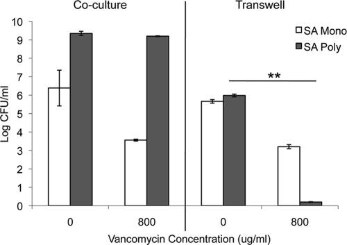
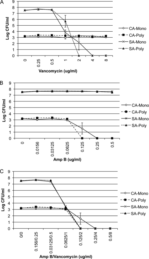
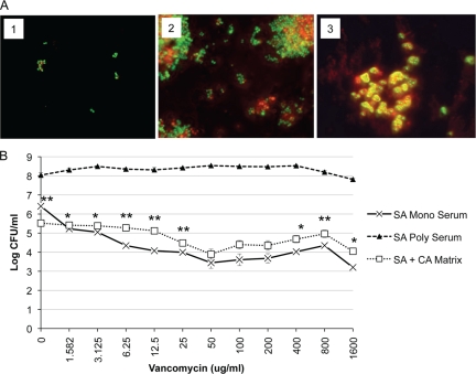
Candida albicans readily forms biofilms on the surface on indwelling medical devices, and these biofilms serve as a source of local and systemic infections. It is estimated that 27% of nosocomial C. albicans bloodstream infections are polymicrobial, with Staphylococcus aureus as the third most common organism isolated in conjunction with C. albicans. We tested whether S. aureus and C. albicans are able to form a polymicrobial biofilm. Although S. aureus formed poor monoculture biofilms in serum, it formed a substantial polymicrobial biofilm in the presence of C. albicans. In terms of architecture, S. aureus formed microcolonies on the surface of the biofilm, with C. albicans serving as the underlying scaffolding. In addition, S. aureus matrix staining revealed a different phenotype in polymicrobial versus monomicrobial biofilms, suggesting that S. aureus may become coated in the matrix secreted by C. albicans. S. aureus resistance to vancomycin was enhanced within the polymicrobial biofilm, required viable C. albicans, and was in part mediated by C. albicans matrix. However, the growth or sensitivity to amphotericin B of C. albicans is not altered in the polymicrobial biofilm.


Figures

FIG. 1.
FIG. 1.
S. aureus biofilm growth in serum and BHI. S. aureus strain ATCC 29523 was grown overnight at 37°C in BHI broth. Cells were washed, counted and diluted into different media: bovine serum (BS), heat-inactivated bovine serum (BS HI), or BHI. (A) For biofilm formation, cultures were diluted to 107 CFU/ml, and 300-μl aliquots were added to tissue culture-treated chamber slides and incubated at 37°C for 24 h without shaking. Biofilm formation was monitored by fluorescence microscopy and SEM. (Panels 1 to 3) For fluorescence microscopy, biofilms were stained with SYTO 9 (viable microbial cells stain green) and ConA-Texas Red (extracellular matrix stains red). SA, S. aureus; Mono, monomicrobial biofilm. (Panels 4 to 6) For SEM, biofilms formed on chamber slides were processed for SEM, cut, and mounted into stubs. Magnifications, ×600 (panels 1 to 3) and ×5,000 (panels 4 to 6). Bars, 10 μm. (B) For biofilm formation, cultures were diluted to 106 CFU/ml, and 100-μl aliquots were added to tissue culture-treated 96-well plates and incubated at 37°C for 24 h without shaking. Biofilm formation was monitored by the CFU assay. Washed plates were washed after 1 h to remove nonadherent cells, while unwashed plates were left alone. Biofilms were disrupted by sonication, and the number of CFU was determined by plating. Experiments were performed in duplicate, and the results are averages of two separate trials.
FIG. 2.
FIG. 2.
Polymicrobial C. albicans-S. aureus biofilm formation in serum. C. albicans SC5314 was grown overnight in SDB at 30°C. S. aureus ATCC 29523 was grown overnight at 37°C in BHI. Both species were washed, counted, and diluted in different media. C. albicans (106 CFU/ml) and S. aureus (107 CFU/ml) were concurrently added to 96-well tissue culture-treated chamber slides and incubated for 24 h at 37°C. Biofilm formation was monitored by fluorescence microscopy and SEM (A) and CFU analysis (B). (A, panels 1 to 3) For fluorescence microscopy, biofilms were stained with calcofluor white (blue stain and blue arrows show fungal cell wall), SYTO 9 (green stain and green arrows show viable microbial cells), and ConA-Texas Red (red stain and red arrows show extracellular matrix). CA, C. albicans; Mono, monomicrobial biofilm; CA/SA Poly, C. albicans and S. aureus polymicrobial biofilm. (Panels 4 to 6) For SEM, biofilms formed on chamber slides were processed for SEM, cut, and mounted into stubs. Magnifications, ×200 (panels 1 and 2), ×1,000 (panel 3), and ×2,000 (panels 4 to 6). Bars, 20 μm. (B) For the CFU assay, plates were washed to remove nonadherent cells. Biofilms were disrupted by sonication, and the number of CFU was determined by plating onto selective media. Experiments were performed in sextuplicate. The data represent the averages of four experiments. Values that were significantly different (P < 0.01) for the S. aureus monomicrobial biofilm versus the S. aureus polymicrobial biofilm are indicated by the bars and asterisks.
FIG. 3.
FIG. 3.
Effect of polymicrobial biofilm formation on antimicrobial drug resistance. Polymicrobial biofilms were formed as described in the legend to Fig. 4 using C. albicans SC5314 (106 CFU/ml) and S. aureus ATCC 29523 (107 CFU/ml) added concurrently to 96-well tissue culture-treated plates in 50% BS. Various concentrations (in micrograms per milliliter) of vancomycin alone (A), amphotericin B alone (B), or a combination of both drugs (C) were tested. After incubation for 24 h at 37°C, the number of CFU was determined by plating onto selective media. Experiments were performed in duplicate, and the data represent the averages of two independent experiments. CA, C. albicans; SA, S. aureus; Mono, monomicrobial biofilm; Poly, polymicrobial biofilm.
FIG. 4.
FIG. 4.
Effect of separation of S. aureus and C. albicans on biofilm formation and vancomycin resistance using a Transwell system. C. albicans (106 CFU/ml) and S. aureus (107 CFU/ml) were added to either 24-well tissue culture-treated plates or 0.4-μm Transwell plates and incubated for 24 h at 37°C. For Transwell plates, S. aureus was added to the bottom of the well, and C. albicans was added to the top of the membrane to allow passage of secreted factors. Vancomycin (800 μg/ml) was added to the bottom well, and plates were incubated an additional 24 h at 37°C. The plates were washed to remove nonadherent cells, and growth was monitored by the CFU assay. Experiments were performed in duplicate, and the data represent the averages of two independent experiments. Values that were significantly different (P < 0.01) for S. aureus in a polymicrobial biofilm with no drug versus S. aureus in a polymicrobial biofilm with 800 μg/ml vancomycin are indicated by the bar and asterisks. SA, S. aureus; Mono, monomicrobial biofilm; Poly, polymicrobial biofilm.
FIG. 5.
FIG. 5.
Effect of planktonic polymicrobial growth on antimicrobial drug resistance. C. albicans SC5314 (103 CFU/ml) alone, S. aureus ATCC 25923 (103 CFU/ml) alone, or C. albicans (103 CFU/ml) and S. aureus (103 CFU/ml) together were added to 96-well, deep-well polypropylene plates in 50% BS. Amp B alone, vancomycin alone, or Amp B and vancomycin combined were added to the wells. The plates were incubated at 35°C in an orbital shaker at 175 rpm for 18 h, and fungal and bacterial viability was monitored by the CFU assay. Experiments were performed in duplicate, and data represent averages of two independent experiments. CA, C. albicans; SA, S. aureus; Mono, monomicrobial biofilm; Poly, polymicrobial biofilm.
FIG. 6.
FIG. 6.
Effect of C. albicans biofilm matrix on S. aureus vancomycin resistance. C. albicans biofilm matrix was isolated as described in Materials and Methods. (A) S. aureus (107 CFU/ml) was added to chamber slides in 50% BS (panel 1), BHI (panel 2), or C. albicans biofilm matrix (panel 3). After 24 h, cells were stained with SYTO 9 (viable microbial cells stain green) and ConA-Texas Red (extracellular matrix stain red). (B) S. aureus (107 CFU/ml) was added alone, with C. albicans biofilm matrix, or with C. albicans (106 CFU/ml) to 96-well tissue culture-treated plates in 50% BS. After incubation for 24 h at 37°C, the plates were washed, and vancomycin was added. The plates were incubated for an additional 24 h. The plates were washed to remove nonadherent cells, and growth was monitored by the CFU assay. Experiments were performed in duplicate, and results are representative of two independent experiments. Values that were significantly different (P < 0.01) for S. aureus monomicrobial biofilm in the presence of serum versus S. aureus in the presence of C. albicans matrix are indicated (**). CA, C. albicans; SA, S. aureus; Mono, monomicrobial biofilm; Poly, polymicrobial biofilm.

References

    1. Adam, B., G. S. Baillie, and L. J. Douglas. 2002. Mixed species biofilms of Candida albicans and Staphylococcus epidermidis. J. Med. Microbiol. 51:344-349. - PubMed
    1. Al-Fattani, M. A., and L. J. Douglas. 2006. Biofilm matrix of Candida albicans and Candida tropicalis: chemical composition and role in drug resistance. J. Med. Microbiol. 55:999-1008. - PubMed
    1. Al-Fattani, M. A., and L. J. Douglas. 2004. Penetration of Candida biofilms by antifungal agents. Antimicrob. Agents Chemother. 48:3291-3297. - PMC - PubMed
    1. Baillie, G. S., and L. J. Douglas. 2000. Matrix polymers of Candida biofilms and their possible role in biofilm resistance to antifungal agents. J. Antimicrob. Chemother. 46:397-403. - PubMed
    1. Carlson, E. 1983. Effect of strain of Staphylococcus aureus on synergism with Candida albicans resulting in mouse mortality and morbidity. Infect. Immun. 42:285-292. - PMC - PubMed

Publication types

MeSH terms

LinkOut - more resources