. 2009 Sep;77(9):4081-91.
doi: 10.1128/IAI.00320-09. Epub 2009 Jun 29.

LuxS promotes biofilm maturation and persistence of nontypeable haemophilus influenzae in vivo via modulation of lipooligosaccharides on the bacterial surface

Affiliations

LuxS promotes biofilm maturation and persistence of nontypeable haemophilus influenzae in vivo via modulation of lipooligosaccharides on the bacterial surface

Chelsie E Armbruster et al. Infect Immun. 2009 Sep.

Abstract

Nontypeable Haemophilus influenzae (NTHI) is an extremely common airway commensal which can cause opportunistic infections that are usually localized to airway mucosal surfaces. During many of these infections, NTHI forms biofilm communities that promote persistence in vivo. For many bacterial species, density-dependent quorum-signaling networks can affect biofilm formation and/or maturation. Mutation of luxS, a determinant of the autoinducer 2 (AI-2) quorum signal pathway, increases NTHI virulence in the chinchilla model for otitis media infections. For example, bacterial counts in middle-ear fluids and the severity of the host inflammatory response were increased in luxS mutants compared with parental strains. As these phenotypes are consistent with those that we have observed for biofilm-defective NTHI mutants, we hypothesized that luxS may affect NTHI biofilms. A luxS mutant was generated using the well-characterized NTHI 86-028NP strain and tested to determine the effects of the mutation on biofilm phenotypes in vitro and bacterial persistence and disease severity during experimental otitis media. Quantitation of the biofilm structure by confocal microscopy and COMSTAT analysis revealed significantly reduced biomass for NTHI 86-028NP luxS biofilms, which was restored by a soluble mediator in NTHI 86-028NP supernatants. Analysis of lipooligosaccharide moieties using an enzyme-linked immunosorbent assay and immunoblotting showed decreased levels of biofilm-associated glycoforms in the NTHI 86-028NP luxS strain. Infection studies showed that NTHI 86-028NP luxS had a significant persistence defect in vivo during chronic otitis media infection. Based on these data, we concluded that a luxS-dependent soluble mediator modulates the composition of the NTHI lipooligosaccharides, resulting in effects on biofilm maturation and bacterial persistence in vivo.


Figures
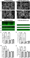
FIG. 1.
FIG. 1.
Microscopic analysis of in vitro biofilm formation in flow chambers. NTHI strains expressing gfp were cultured in flow cells under continuous-medium-flow conditions for 72 h. (A to D) SEM of biofilms formed by NTHI 86-028NP and NTHI 86-028NP luxS at magnifications of ×5,000 (A and B) and ×10,000 (C and D). (E) CLSM vertical z-series images of biofilms formed by NTHI 86-028NP and NTHI 86-028NP luxS at 24 h, 48 h, and 72 h. (F to I) Biofilm measurements for NTHI 86-028NP (open bars) and NTHI 86-028NP luxS (gray bars) obtained using COMSTAT, including measurements of total biomass (F), average thickness (G), roughness (H), and surface/volume ratio (I). Significance was determined by a two-way ANOVA with a post hoc test of significance (***, P ≤ 0.001; **, P ≤ 0.01; *, P ≤ 0.05). The error bars indicate standard errors of the means.
FIG. 2.
FIG. 2.
Complementation of luxS with exogenous AI-2. NTHI strains expressing gfp were separated by 0.2-μm Anapore membranes and cultured in two-well chamber slides under stationary conditions for 12 h. Biofilm measurements of total biomass (A) and average thickness (B) were obtained from CLSM vertical z-series images using COMSTAT. Significance was determined by a nonparametric t test (***, P ≤ 0.001; *, P ≤ 0.05). The error bars indicate standard errors of the means.
FIG. 3.
FIG. 3.
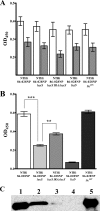
Analysis of LOS composition. Modified whole-bacterium ELISA using treatment with 80 mU neuraminidase as a negative control for reactivity with L. flavus lectin for detection of surface sialic acid (A) (n = 2) and ELISA using anti-PCho monoclonal antibody for detection of surface phosphorylcholine (B) (n = 5). Significance was determined by a nonparametric t test (***, P ≤ 0.001; **, P ≤ 0.01). The error bars indicate standard errors of the means. (C) Western blot of purified LOS for detection of total PCho from NTHI 86-028NP (lane 1), NTHI 86-028NP luxS IRA::luxS (lane 2), NTHI 86-028NP luxS (lane 3), NTHI 86-028NP licD (lane 4), and NTHI 86-028NP licON (lane 5).
FIG. 4.
FIG. 4.
luxS promotes biofilm formation and persistence in the chinchilla model of OM. (A) Middle ear of an uninfected control animal at 21 days postinfection. (B to H) Biofilms in the middle ear visualized at 7 days (B and E), 14 days (C and F), and 21 days (D, G, and H) postinfection. Panels B, C, and D show biofilms from animals infected with NTHI 86-028NP, and panels E, F, G, and H show biofilms from animals infected with NTHI 86-028NP luxS. Panel G is representative of five of six ears infected with NTHI 86-028NP luxS at 21 days postinfection, while panel H shows the only ear containing a biofilm at this time point. (I and J) Bacterial counts for middle-ear effusion and homogenized bullae after infection with NTHI 86-028NP (filled symbols) or NTHI 86-028NP luxS (open symbols). The limit of detection was 30 CFU/ear. Statistical significance was determined by two-way ANOVA with a post hoc test of significance. The horizontal bars indicate median values.
FIG. 5.
FIG. 5.
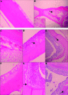
Hematoxylin and eosin staining of bullar sections. (A) PBS control (magnification, ×20) showing smooth bone surface with few neutrophils and no significant evidence of tissue injury. (B) Focal osteomyelitis (arrow) 14 days after inoculation with NTHI 86-028NP. The marrow space has been effaced by intense inflammation, as indicated by comparison to the normal bone marrow on the left. Magnification, ×2. (Inset) Higher magnification (×40) of the exudate. (C to E) Stained sections from animals at 7, 14, and 21 days after infection with NTHI 86-028NP. Magnification, ×20. (F to H) Sections from animals at 7, 14, and 21 days after infection with NTHI 86-028NP luxS. Magnification, ×20. The arrows indicate periosteal hyperplasia.

References

    1. Bakaletz, L. O., B. M. Tallan, T. Hoepf, T. F. DeMaria, H. G. Birck, and D. J. Lim. 1988. Frequency of fimbriation of nontypable Haemophilus influenzae and its ability to adhere to chinchilla and human respiratory epithelium. Infect. Immun. 56331-335. - PMC - PubMed
    1. Boles, B. R., and A. R. Horswill. 2008. Agr-mediated dispersal of Staphylococcus aureus biofilms. PLoS Pathog 4e1000052. - PMC - PubMed
    1. Bouchet, V., D. W. Hood, J. Li, J. R. Brisson, G. Randle, A. Martin, Z. Li, R. Goldstein, and E. K. Schweda. 2003. Host-derived sialic acid is incorporated into Haemophilus influenzae lipopolysaccharide and is a major virulence factor in experimental otitis media. Proc. Natl. Acad. Sci. USA 1008898-8903. - PMC - PubMed
    1. Campagnari, A. A., M. R. Gupta, K. C. Dudas, T. F. Murphy, and M. A. Apicella. 1987. Antigenic diversity of lipooligosaccharides of nontypable Haemophilus influenzae. Infect. Immun. 55882-887. - PMC - PubMed
    1. Campagnari, A. A., S. M. Spinola, A. J. Lesse, Y. Abu Kwaik, R. E. Mandrell, and M. Apicella. 1990. Lipooligosaccharide epitopes shared among Gram-negative non-enteric mucosal pathogens. Microb. Pathog. 8353-362. - PubMed

Publication types