. 2009 Jul 14;106(28):11606-11.
doi: 10.1073/pnas.0905165106. Epub 2009 Jun 29.

A site-specific, multiplexed kinase activity assay using stable-isotope dilution and high-resolution mass spectrometry

Affiliations

A site-specific, multiplexed kinase activity assay using stable-isotope dilution and high-resolution mass spectrometry

Yonghao Yu et al. Proc Natl Acad Sci U S A. .

Abstract

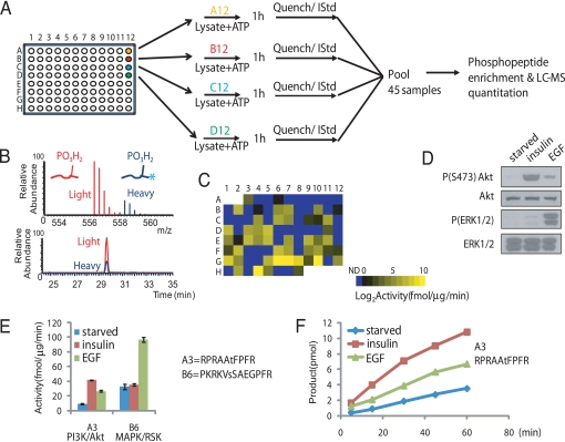
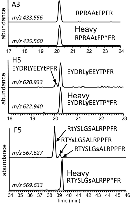
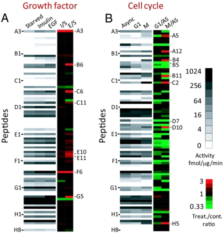
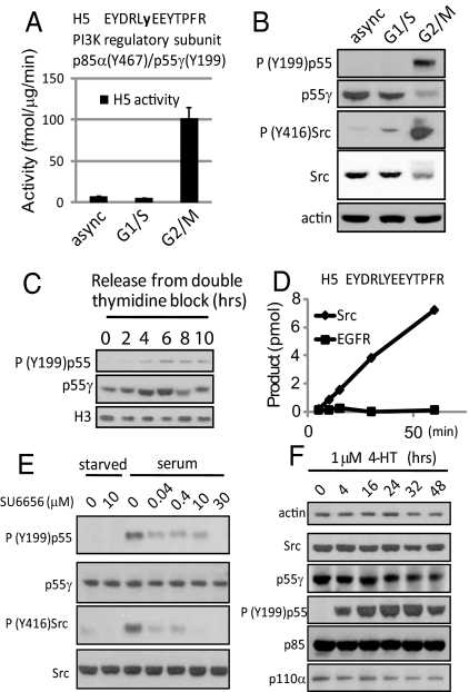
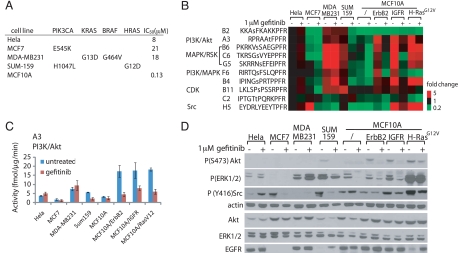
Most kinases are capable of recognizing and phosphorylating peptides containing short, linear sequence motifs. To measure the activation state of many kinases from the same cell lysate, we created a multiplexed, mass-spectrometry-based in vitro kinase assay. Ninety chemically synthesized peptides derived from well-characterized peptide substrates and in vivo phosphorylation sites with either known or previously unidentified upstream kinases were reacted individually in a plate format with crude cell lysates and ATP. Phosphorylation rates were directly measured based on the addition of 90 same-sequence, site-specific phosphopeptides enriched in stable isotopes to act as ideal quantitative internal standards for analysis by liquid chromatography coupled to tandem mass spectrometry. This approach concurrently measured up to 90 site-specific peptide phosphorylation rates, reporting a diagnostic fingerprint for activated kinase pathways. We applied this unique kinome-activity profiling strategy in a variety of cellular settings, including mitogen stimulation, cell cycle, pharmacological inhibition of pathways, and to a panel of breast cancer cell lines. Finally, we identified the source of activity for a peptide (derived from a PI3K regulatory subunit) from our library. This peptide substrate demonstrated mitotic and tyrosine-specific phosphorylation, which was confirmed to be a novel Src family kinase site in vivo.


Conflict of interest statement

The authors declare no conflict of interest.

Figures

Fig. 1.
Fig. 1.
General scheme of the KAYAK strategy. (A) Overview of the procedure, where 90 synthetic peptides are used as substrates for in vitro kinase assays. (B) Example of high-resolution MS and elution chromatogram for a light and heavy (internal standard; IStd) pair of phosphopeptides. Asterisk indicates incorporation of a proline residue containing 6 additional Daltons of heavy isotopes. (C) Intensity map representation of substrate phosphorylation activities (average of triplicate experiments) from 6 μg starved HEK293 cell lysates toward each of the 90 peptides in their respective plate positions. ND, not detected, indicates the ones below the threshold. (D) Immunoblot analysis of the lysates of insulin- and EGF-stimulated HEK293 cells using the indicated antibodies. (E) Examples of phosphorylation rates (performed from 3 separate experiments) for a preferred Akt (A3; RPRAAtFPFR) and RSK (B6; PKRKVsSAEGPFR) substrate, respectively, using the lysates in (D). (F) Time course of the Akt-specific substrate, A3, phosphorylation reaction using the same lysates in D.
Fig. 2.
Fig. 2.
Site-specific measurement of peptide phosphorylation rates. Peptides phosphorylated at different Ser/Thr/Tyr residues were resolved by LC and phosphorylation sites were localized by concurrent tandem MS analysis (see Fig. S3 for an example). Perfect co-elution of the internal standard and product facilitated the determination of a site-specific phosphorylation rate. Extracted ion chromatograms were produced using a ± 10 ppm tolerance surrounding the predicted mass-to-charge (m/z) ratio for each phosphopeptide and each internal standard (contains heavy proline residue, indicated by asterisk). Confirmed phosphorylation sites are bold and lowercase. Peptide A3 has a single acceptor site residue, and its phosphorylation product perfectly co-eluted with the internal standard. In contrast, the phosphorylation products of H5 and F5 both contained multiple position isomers. In the case of F5, no site-specific phosphorylation toward the site in the IStd was detected.
Fig. 3.
Fig. 3.
Peptide phosphorylation rates accurately report pathway activation states. (A) Intensity map of kinase activities of starved (S), insulin-stimulated (I), and EGF-stimulated (E) HEK293 cells (average of triplicate experiments). Fold change over the starved state is shown. Peptides with signals below detection threshold were not included for ratio calculation. (B) Intensity map of kinase activities of asynchronously growing (AS) HeLa cells, and cells arrested in either G1/S or G2/M phase of the cell cycle. Fold change over the asynchronous state is shown. Peptides were ordered according to their positions in the 96-well plate. Sequences of all peptides can be found in Table S1.
Fig. 4.
Fig. 4.
Identification and validation of Src family kinase activity toward Tyr-199 of PI3K regulatory subunit p55. (A) Activity toward substrate peptide H5 using lysates of asynchronously growing HeLa cells and cells arrested in G1/S and G2/M phase. (B) Immunoblot analysis of phospho-PI3K regulatory subunit p55 (Tyr-199) levels in these same lysates. (C) Phospho-PI3K regulatory subunit p55 (Tyr-199) immunoreactivity in HeLa cells undergoing a natural mitosis. (D) In vitro phosphorylation of peptide H5 using 2 ng of purified Src or EGFR. (E) Treatment of asynchronously growing HEK293 cells using a Src family kinase (SFK) specific inhibitor, SU6656. (F) Immunoblot analysis of vSrc-ER expressing MCF10A cells treated with 1 μM 4-HT to activate vSrc.
Fig. 5.
Fig. 5.
Peptide phosphorylation rates accurately report activating mutations in kinase pathways. Seven breast cancer cell lines and HeLa cells were examined. (A) Summary of the activating mutations within the PI3K and MAPK pathways and IC50 of these cells toward gefitinib treatment (22, 25, 26). (B) KAYAK activities (average of duplicates, shown as fold difference normalized to asynchronously growing HeLa cells) in cancer cell lines with or without 1 μM treatment of gefitinib for 24 h. Peptide sequences with their reported kinases pathways/kinases are shown. (C) Example of activity profile from (B) for A3 peptide (PI3K/Akt pathway) across these cell lines. (D) Western blot analysis of the lysates for the cell lines using the indicated antibodies.

References

    1. Hanahan D, Weinberg RA. The hallmarks of cancer. Cell. 2000;100:57–70. - PubMed
    1. McLendon R, et al. Comprehensive genomic characterization defines human glioblastoma genes and core pathways. Nature. 2008;455:1061–1068. - PMC - PubMed
    1. Ren R. Mechanisms of BCR-ABL in the pathogenesis of chronic myelogenous leukaemia. Nat Rev Cancer. 2005;5:172–183. - PubMed
    1. Yeatman TJ. A renaissance for SRC. Nat Rev Cancer. 2004;4:470–480. - PubMed
    1. Irish JM, et al. Single cell profiling of potentiated phospho-protein networks in cancer cells. Cell. 2004;118:217–228. - PubMed

Publication types