. 2009 Jul 14;106(28):11617-22.
doi: 10.1073/pnas.0903768106. Epub 2009 Jun 29.

A "latent niche" mechanism for tumor initiation

Affiliations

A "latent niche" mechanism for tumor initiation

Marie McGovern et al. Proc Natl Acad Sci U S A. .

Abstract

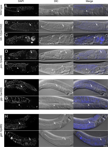
Stem cells, their niches, and their relationship to cancer are under intense investigation. Because tumors and metastases acquire self-renewing capacity, mechanisms for their establishment may involve cell-cell interactions similar to those between stem cells and stem cell niches. On the basis of our studies in Caenorhabditis elegans, we introduce the concept of a "latent niche" as a differentiated cell type that does not normally contact stem cells nor act as a niche but that can, under certain conditions, promote the ectopic self-renewal, proliferation, or survival of competent cells that it inappropriately contacts. Here, we show that ectopic germ-line stem cell proliferation in C. elegans is driven by a latent niche mechanism and that the molecular basis for this mechanism is inappropriate Notch activation. Furthermore, we show that continuous Notch signaling is required to maintain ectopic germ-line proliferation. We highlight the latent niche concept by distinguishing it from a normal stem cell niche, a premetastatic niche and an ectopic niche. One of the important distinguishing features of this mechanism for tumor initiation is that it could operate in the absence of genetic changes to the tumor cell or the tumor-promoting cell. We propose that a latent niche mechanism may underlie tumorigenesis and metastasis in humans.


Conflict of interest statement

The authors declare no conflict of interest.

Figures

Fig. 1.
Fig. 1.
“Latent niche” mechanism of tumor initiation. (A) Schematic representation of the cellular mechanism by which a delay in germ-line differentiation causes tumorous growth in the Caenorhabditis elegans proximal germ line in response to a latent niche. During gonadogenesis, the distance between the distal tip cell (DTC) (red) and the proximal gonad depends on both an intrinsic DTC migration program and the number of germ cells in the gonad (at each time point depicted, distal is to the left and proximal is to the right). In the wild type, proximal germ cells enter meiosis in the middle of the third larval stage (mid-L3), and the proximal sheath cells (blue) are born in the mid-L4. Only the two proximal-most pairs of sheath cells, Sh4 and Sh5, are depicted here. Undifferentiated and differentiated germ cells are indicated in yellow and green, respectively. Two mutant conditions display insufficient early gonad arm extension: hlh-12(Pro/Tum) displays a DTC-autonomous migration defect, and pro-1(Pro) displays a sheath-lineage-autonomous germ-line proliferation defect. The designation hlh-12(Pro/Tum) refers to the observation that hlh-12 mutant individuals with smaller tumors did not contain gametes between the distal and the proximal proliferation centers (“Tum”), whereas those with larger tumors often contained gametes in the center (“Pro”). A third mutant condition, glp-1(Pro), displays a germ-line-autonomous delay in differentiation. In contrast to the wild type, in each mutant undifferentiated germ cells contact the proximal sheath cells (latent niche) in the L4, resulting in a proximal tumor in the adult. Black arrows indicate time, and small blue arrows indicate DSL ligand signaling. (B) (Left and Center) Expression of nuclear-localized Papx-1::NLSlacZ (21) and Papx-1::GFP (naEx156) in a subset of proximal gonadal sheath and spermathecal cells (fixed and live worms, respectively). (Right) Expression of Parg-1::GFP (naEx180) in the proximal gonadal sheath. Proximal sheath pairs 4 and 5 (Sh4 and Sh5) are indicated by dotted-line and solid-line arrows, respectively. Arrowheads point to cells of the distal spermatheca. Expression from Papx-1::lacZ was observed in 59%, 97%, and 100% of animals in Sh4, Sh5, and spermatheca, respectively (n = 88). Expression from Papx-1::GFP was observed in 78%, 100%, and 100%, in Sh4, Sh5, and spermatheca, respectively (n = 56). We observed a more dynamic gonad pattern for Parg-1::GFP. Expression was not observed in L2 or L3 gonads (n = 12) and first appears in the sheath cells in the mid-L4, in Sh4 and Sh5 (12/12 gonad arms), and occasionally also in Sh3 (1/12). By the late L4, Sh4 and Sh5 continue to express Parg-1::GFP (6/6) with 2/6 also expressing the transgene in Sh1–3. In very young adults (before embryogenesis), expression remained in Sh4 and Sh5 in all animals but was less frequently observed in Sh1–3 (2/9 gonad arms). Finally, in adults bearing embryos (n = 48 gonad arms), 25% showed no expression. Expression was observed in 73% of Sh4–5, and 15% of these also showed expression in Sh3, whereas 2% showed more distal sheath expression. [Scale bars, 20 μm.]
Fig. 2.
Fig. 2.
Suppression of proximal germ-line tumors by L1 RNAi feeding. (A–I) Representative gonad arms in fixed DAPI-stained worms. (A) Wild type grown on control RNAi bacteria (carrying the L4440 empty-vector plasmid). For the remaining images, each pair of panels includes control on the top (B, D, F, H) and apx-1(RNAi) on the bottom (C, E, G, I): (B, C) hlh-12(ok1532); (D, E) pro-1(na48); (F, G) glp-1(ar202); (H, I) gld-1(q485). hlh-12 encodes a basic helix–loop–helix transcription factor (45), pro-1 encodes an ortholog of yeast IPI3 (15), and gld-1 encodes a KH-domain protein (46). Arrows and arrowheads indicate undifferentiated and differentiated germ cells in the proximal oviduct, respectively. See Table 1 for quantification. [Scale bars, 20 μm.]
Fig. 3.
Fig. 3.
Control of proximal tumors. (A) LAG-2 can replace APX-1 in the latent niche role. Gonad arms from live worms raised on the indicated RNAi bacteria. In each RNAi treatment, siblings were scored from mothers of the genotype pro-1(na48); naEx152[Papx-1::lag-2]. Control is L4440 RNAi; graph shows quantification. (B) Suppression of adult tumors by reducing ligand or receptor activity. (Upper, day 1) Gonad arms from representative early adult pro-1(na48) animals raised to early adulthood on control L4440 RNAi. (Lower, day 2) Same individual gonad arm in the panel above after an additional day of feeding on bacteria inducing the indicated RNAi. Arrows indicate undifferentiated germ cells; arrowheads indicate differentiated germ cells. See Table 1 for quantification. [Scale bars in A and B, 20 μm.] (C) Generalized latent niche mechanism. (Row 1) Normal conditions: latent niche mechanism inactive. Cell–cell contacts between the latent niche cell (blue) and its unresponsive neighbors (green) do not cause inappropriate self-renewal or proliferation. Although the latent niche is producing a potential niche-like renewal-promoting signal, under normal conditions, neighboring cells do not respond to this signal. (Rows 2 and 3) Tumor initiation: latent niche mechanism active. Several different scenarios could lead to activation of the latent niche mechanism, two of which are schematized here. (Row 2) A delay in differentiation before the appearance of the latent niche ultimately juxtaposes the latent niche with proliferation-competent cells (yellow); (Row 3) anatomical changes or the migration of potentially self-renewing tumor cells juxtaposes proliferation-competent cells with a resident latent niche. Time proceeds from left to right. Dotted lines indicate the positions of cells not yet born or newly opened vacancies.

References

    1. Hubbard EJ. Caenorhabditis elegans germ line: A model for stem cell biology. Dev Dyn. 2007;236:3343–3357. - PMC - PubMed
    1. Kimble J, Crittenden SL. Controls of germline stem cells, entry into meiosis, and the sperm/oocyte decision in Caenorhabditis elegans. Annu Rev Cell Dev Biol. 2007;23:405–433. - PubMed
    1. Hansen D, Schedl T. The regulatory network controlling the proliferation-meiotic entry decision in the Caenorhabditis elegans germ line. Curr Top Dev Biol. 2006;76:185–215. - PubMed
    1. Lambie EJ, Kimble J. Two homologous regulatory genes, lin-12 and glp-1, have overlapping functions. Development. 1991;112:231–240. - PubMed
    1. Henderson S, Gao D, Lambie E, Kimble J. lag-2 may encode a signaling ligand for the GLP-1 and LIN-12 receptors of C. elegans. Development. 1994;120:2913–2924. - PubMed

Publication types

Substances