. 2009 Jun 30;7(6):e1000140.
doi: 10.1371/journal.pbio.1000140. Epub 2009 Jun 30.

Attention enhances the retrieval and stability of visuospatial and olfactory representations in the dorsal hippocampus

Affiliations

Attention enhances the retrieval and stability of visuospatial and olfactory representations in the dorsal hippocampus

Isabel A Muzzio et al. PLoS Biol. .

Erratum in

  • PLoS Biol. 2010;8(10). doi: 10.1371/annotation/5e28240c-6186-43eb-b319-79c391d9468a
  • PLoS Biol. 2010;8(10). doi: 10.1371/annotation/d0e5ef6f-d08b-474a-8fde-aeebeee7369d

Abstract

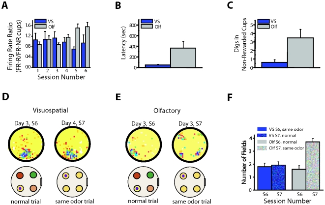
A key question in the analysis of hippocampal memory relates to how attention modulates the encoding and long-term retrieval of spatial and nonspatial representations in this region. To address this question, we recorded from single cells over a period of 5 days in the CA1 region of the dorsal hippocampus while mice acquired one of two goal-oriented tasks. These tasks required the animals to find a hidden food reward by attending to either the visuospatial environment or a particular odor presented in shifting spatial locations. Attention to the visuospatial environment increased the stability of visuospatial representations and phase locking to gamma oscillations--a form of neuronal synchronization thought to underlie the attentional mechanism necessary for processing task-relevant information. Attention to a spatially shifting olfactory cue compromised the stability of place fields and increased the stability of reward-associated odor representations, which were most consistently retrieved during periods of sniffing and digging when animals were restricted to the cup locations. Together, these results suggest that attention selectively modulates the encoding and retrieval of hippocampal representations by enhancing physiological responses to task-relevant information.


Conflict of interest statement

The authors have declared that no competing interests exist.

Figures

Figure 1
Figure 1. Experimental design and task acquisition.
(A) Recording location. Schematic diagram of tetrode placements in the left dorsal hippocampus CA1 pyramidal cell layer (red circles). (B and C) Goal-oriented tasks. Two groups of mice were trained to find a hidden food reward buried inside one of four cups, which were filled with odor-scented bedding. (B) In the visuospatial task, the location of the reward remained fixed throughout training but the scented bedding covering the reward changed from trial to trial. (C) In the olfactory task, the location of the reward shifted from trial to trial in a pseudo-random fashion, but the scented bedding covering the reward remained constant. Odors used were cumin (cu), cinnamon (cinn), cloves (cl), and ginger (gin); the black dot placed on top of one of the cups represents the hidden food reward. (D and E) Task acquisition was equivalent in both groups as illustrated by the similar reduction in (D) latency to find the reward and (E) number of errors [latency: F(5,88) = 22.71, p<0.001; errors: F(5,88) = 12.48, p<0.001], with no significant difference between the two groups in the rate of acquisition [latency: group: F(1,88) = 0.07, p = 0.78; interaction: F(5,88) = 1.07, p = 0.38; errors: group: F(1,84) = 0.25, p = 0.63; interaction: F(5,84) = 0.78, p = 0.57]. Blue: visuospatial group: n = 12; Gray: olfactory group: n = 11; Line plots show session mean±SEM.
Figure 2
Figure 2. Attention to the visuospatial environment enhances place field stability.
Color-coded rate maps showing firing activity of two single CA1 pyramidal cells over three sequential days in animals trained in the visuospatial group. Below each rate map, a cartoon of the arena marks the position of the reward with a red circle. The four waveforms on the right represent a tetrode recording from a single cell. The constancy of the waveforms throughout the three days of training demonstrates recording stability. On day 1, both cells (A and B) displayed unstable and disorganized place fields. As animals learned to attend to the visuospatial environment, the stability and organization of the fields was significantly enhanced. This effect was evident during training and probe trials (T0). Color map indicates neuronal level of activity. White pixels are regions that the animal never visited. Yellow pixels are regions the animal visited but the cell never fired. Orange, red, green, blue, and purple pixels progressively encode higher firing rates that are auto-scaled relative to the peak firing frequency (shown above each rate map).
Figure 3
Figure 3. Attention to a spatially shifting olfactory cue compromises place field stability.
Color-coded rate maps showing firing activity of CA1 pyramidal cells over three sequential days in animals trained in the olfactory group. Color maps, cartoon notations, and waveforms represent the same parameters shown in Figure 2. (A) Olfactory group, cell type 1: A general characteristic of this cell type was the emergence of multiple fields with one often locked to the reward-associated odor. These cells became highly disorganized with successive trials. This effect was evident during training as well as during the probe trial (T0). (B) Olfactory group, cell type 2: In these cells location-specific firing disintegrated quickly. As training progressed, the firing fields of these cells coincided with the location of the reward-associated odor.
Figure 4
Figure 4. During navigation, attention modulates the stability of spatial representations during task performance and free exploration.
(A) Long-term place field stability was analyzed by correlating neuronal activity between the first training trials of each session. Before task acquisition, both groups, visuospatial and olfactory, showed similar levels of stability. After animals learned to attend to the relevant rule, stability was significantly enhanced in animals in the visuospatial group, remaining low after only one training session in the olfactory group. Groups: F(1,63) = 26.93, p<0.001; session: F(4, 63) = 5.49, p<0.001; interaction: F(4,63) = 3.15, p<0.02, groups were different on sessions 2, 3, 4, 5, and 6, p<0.05. (B) Short-term stability was calculated by averaging the correlation values between training trials in each session. Learning to attend to the relevant percept significantly enhanced the short-term stability in the visuospatial group and reduced it in the olfactory group [groups: F(1,83) = 15.20, p<0.001; session: F(5,83) = 4.22, p<0.002, groups were different on sessions 2, 3, 4, 5, and 6, p<0.05; interaction: F(5,83) = 3.34, p<0.008]. (C and D) Examples of cluster projections and rate maps of cells recorded from animals in the visuospatial (C) and olfactory (D) groups on days 1–3 demonstrating recording stability. Color maps and waveforms represent the same parameters shown in Figure 2. (E) Correlation coefficients calculated during the probe trials (T0) in sequential sessions revealed enhanced long-term place field stability in animals trained in the visuospatial, but not the olfactory group [group: F(1,97) = 22.26, p<0.001; session: F(6,97) = 2.49, p<0.03; interaction: F(6,97) = 2.58, p<0.03]. Post hoc analysis showed that the groups were significantly different on session 3, 4, 5, 6, and 7 (p<0.02), but not on session 1 or 2 before the animals learned the task (p>0.05). Line plots show session mean±SEM. BL, baseline.
Figure 5
Figure 5. Attention to a task-relevant olfactory cue enhances retrieval of a reward-associated odor.
(A) Firing rate responses to the reward-associated odor increased during periods of sniffing and digging only in the olfactory group. (B–F) Same odor trial: During this trial the four cups in the arena contained the same scented bedding, but the reward was hidden in only one of the cups. (B and C) The same odor trial only affected the behavior of olfactory animals as illustrated in the increase in the latency to find the reward (B) and the number of digs in the non-rewarded cups (C). (D and E) Rate maps of animals trained in the visuospatial (D) and olfactory (E) groups during the same odor trial. (D) The place fields of animals in the visuospatial group were not affected. (E) In the olfactory group, however, the fields that coincided with the location of the reward-associated odor during training broke down into four fields locked to the position of the four cups. Cartoon below each rate map indicates the positions of odors and the buried reward (black dot) on each trial. The color map is the same as that shown in Figure 2. (F) Number of fields associated with the four-cup locations during the last training session [day 3, session 6 (S6)] and the same odor trial [day 4, session 7 (S7)] recorded in the visuospatial and olfactory groups. The number of fields significantly increased during the same odor trial in the olfactory group [t(6) = −3.48, p<0.02], showing no significant difference in the visuospatial group [t(6) = 1, p = .356]. Histograms show trial mean±SEM. Olf, olfactory; S, session; VS, visuospatial, yellow: ginger, green: cumin, red: cinnamon, pink: cloves.
Figure 6
Figure 6. Place fields from animals trained in the visuospatial task are locked to the proximal visuospatial cues in the environment.
(A and B) Behavioral effects of the cue control and cue conflict rotations. During the cue control trial the behavioral performance of both groups was unaffected, as illustrated by the equivalent short latencies to find the reward [A, latency: t(17) = 0.08, p = 0.94] and the low number of errors made prior to obtaining reward [B, errors: t(15) = 0.07, p<0.93]. In contrast, during the cue conflict trial only animals in the visuospatial group were severely disrupted by this manipulation [latency: t(17) = 10.6, p<0.00001; digs in non-rewarded cups: t(17) = 3.87, p<0.0007]. Histograms show trial mean±SEM. (C and D) Color-coded rate maps showing firing activity of CA1 pyramidal cells in response to cue rotations. Cartoon placed below each rate map indicates the position of the reward (red circle) and the direction of rotation of the visuospatial environment (90° counterclockwise (CCW), or clockwise (CW)) in each trial. Color map is the same as that shown in Figure 2. (C) Upper panel: Only cells from animals in the visuospatial group displayed concomitant rotations of the fields when the visuospatial cues in the environment were rotated. Middle and bottom panels: Cue rotations had no effect on the firing pattern of unstable cells from animals in the olfactory group. This was the case for cells that exhibited disorganized firing activity as well as those in which firing activity was locked to the reward-associated odor. (D) Rate maps showing place fields of a stable cell recorded in the olfactory group. Cue control and cue conflict experiments [day 4, session 7, T2 and T3 respectively] did not produce re-mapping of the fields in spite of the physical rotation of the environmental cues. On day 5 (session 8), the cue control and cue conflict experiments were replicated on T1 and T2, and on T3 the animal was tested in the dark with the visuospatial cues covered with black paper. As was the case with the first control trials, the fields remained unchanged. Olf, olfactory; T, trial; VS, visuospatial.
Figure 7
Figure 7. Increase in spike phase locking in animals trained in the visuospatial task.
(A and B) Spike-triggered average (STA) generated using a ±100 ms time window on days 1 (blue line), 2 (red line), and 3 (black line) in a visuospatial (A) and olfactory (B) animal. The STA for the animal in the visuospatial group has a clear oscillatory component with a periodicity of about 20 ms (50 Hz). Such a high-frequency oscillatory component is not seen in the STA of the animal in the olfactory group. (C–F) Relative power of gamma (20–60 Hz) in the ±100 ms STA across sessions. (C) In the visuospatial group the relative power of gamma, which reflects phase locking to gamma oscillations, increases during the first 40 s of each trial being maximal after animals reach asymptotic levels of performance. No increase in relative power is observed in the olfactory group. (D) The increase in relative power observed in the visuospatial group during the initial part of the trial (0–40 s) is not present in the last post-reward segment of the trials (860–900 s). (E) In the olfactory group the relative power of gamma does not change before or after obtaining the reward. (F) A distracter (intermittent flashing lights) decreased phase locking in trials 2, 4, and 6 in comparison to normal trials in which no distracting stimuli were presented. (G and H) Same STA examples as those shown in (A and B) generated with a ±200 ms time window. (I) Relative power of theta (4–12 Hz) in the ±200 ms STAs across sessions. In both groups the relative power of theta is relatively high, consistent with the moderately high power of theta in the LFP (Figure S5C), which is characteristic of hippocampal cells during periods of movement. There were no significant differences between the groups or across training trials. LFP, local field potential; Olf, olfactory; PostR, post-reward; PreR, pre-reward; S, session; VS, visuospatial.

Similar articles

Cited by

References

    1. Ferbinteanu J, Kennedy PJ, Shapiro ML. Episodic memory–from brain to mind. Hippocampus. 2006;16:691–703. - PubMed
    1. Smith DM, Mizumori SJ. Hippocampal place cells, context, and episodic memory. Hippocampus. 2006;16:716–729. - PubMed
    1. Fernandes MA, Moscovitch M, Ziegler M, Grady C. Brain regions associated with successful and unsuccessful retrieval of verbal episodic memory as revealed by divided attention. Neuropsychologia. 2005;43:1115–1127. - PubMed
    1. Schacter DL. Illusory memories: a cognitive neuroscience analysis. Proc Natl Acad Sci U S A. 1996;93:13527–13533. - PMC - PubMed
    1. Neves G, Cooke SF, Bliss TV. Synaptic plasticity, memory and the hippocampus: a neural network approach to causality. Nat Rev Neurosci. 2008;9:65–75. - PubMed

Publication types

MeSH terms