. 2009 Jun 30;4(6):e6093.
doi: 10.1371/journal.pone.0006093.

Synergistic activation of HIV-1 expression by deacetylase inhibitors and prostratin: implications for treatment of latent infection

Affiliations

Synergistic activation of HIV-1 expression by deacetylase inhibitors and prostratin: implications for treatment of latent infection

Sophie Reuse et al. PLoS One. .

Abstract

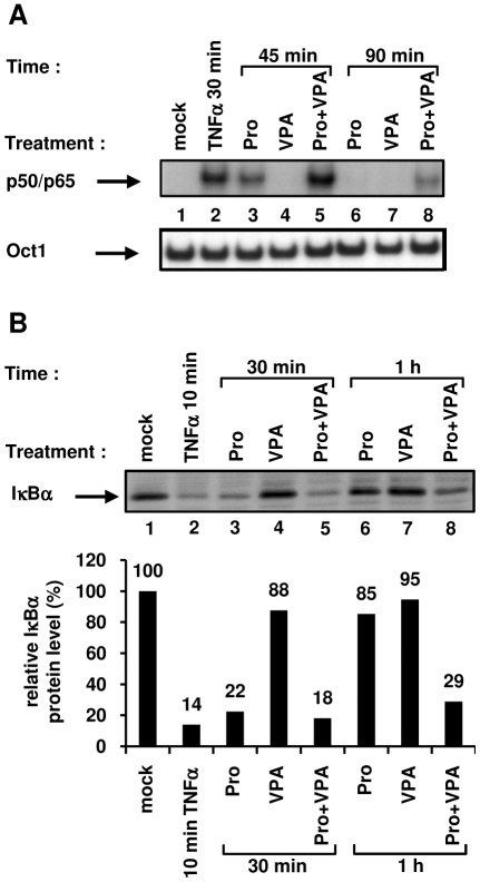
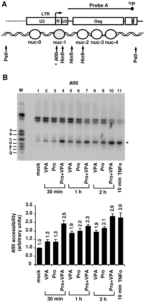
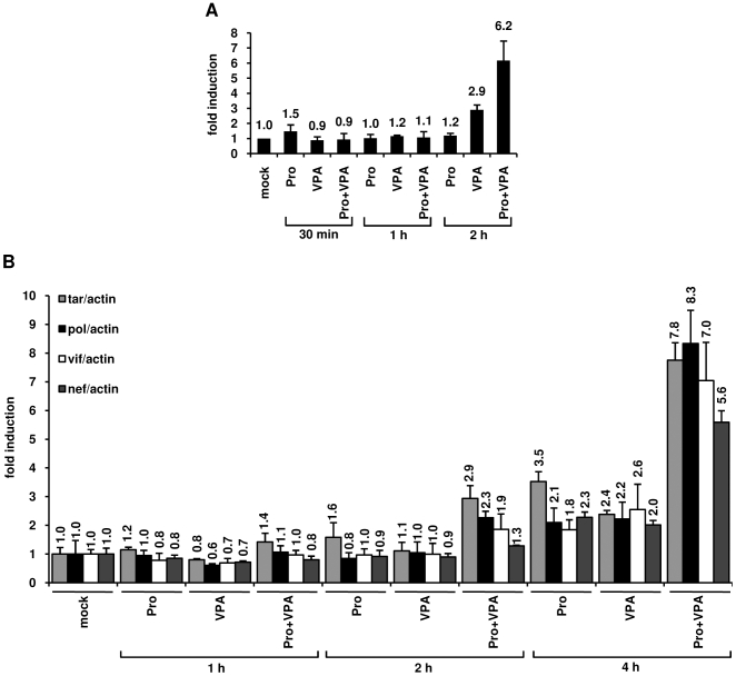
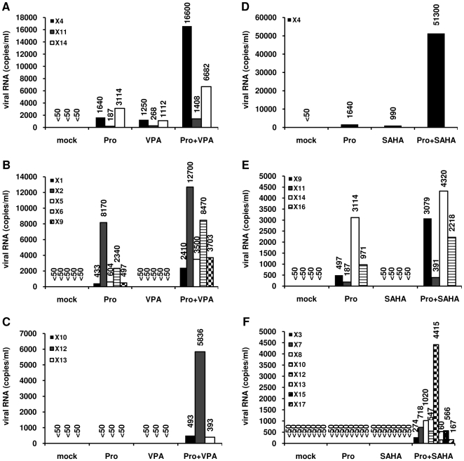
The persistence of transcriptionally silent but replication-competent HIV-1 reservoirs in Highly Active Anti-Retroviral Therapy (HAART)-treated infected individuals, represents a major hurdle to virus eradication. Activation of HIV-1 gene expression in these cells together with an efficient HAART has been proposed as an adjuvant therapy aimed at decreasing the pool of latent viral reservoirs. Using the latently-infected U1 monocytic cell line and latently-infected J-Lat T-cell clones, we here demonstrated a strong synergistic activation of HIV-1 production by clinically used histone deacetylase inhibitors (HDACIs) combined with prostratin, a non-tumor-promoting nuclear factor (NF)- kappaB inducer. In J-Lat cells, we showed that this synergism was due, at least partially, to the synergistic recruitment of unresponsive cells into the expressing cell population. A combination of prostratin+HDACI synergistically activated the 5' Long Terminal Repeat (5'LTR) from HIV-1 Major group subtypes representing the most prevalent viral genetic forms, as shown by transient transfection reporter assays. Mechanistically, HDACIs increased prostratin-induced DNA-binding activity of nuclear NF-kappaB and degradation of cytoplasmic NF-kappaB inhibitor, IkappaBalpha . Moreover, the combined treatment prostratin+HDACI caused a more pronounced nucleosomal remodeling in the U1 viral promoter region than the treatments with the compounds alone. This more pronounced remodeling correlated with a synergistic reactivation of HIV-1 transcription following the combined treatment prostratin+HDACI, as demonstrated by measuring recruitment of RNA polymerase II to the 5'LTR and both initiated and elongated transcripts. The physiological relevance of the prostratin+HDACI synergism was shown in CD8(+)-depleted peripheral blood mononuclear cells from HAART-treated patients with undetectable viral load. Moreover, this combined treatment reactivated viral replication in resting CD4(+) T cells isolated from similar patients. Our results suggest that combinations of different kinds of proviral activators may have important implications for reducing the size of latent HIV-1 reservoirs in HAART-treated patients.


Conflict of interest statement

Competing Interests: The authors have declared that no competing interests exist.

Figures

Figure 1
Figure 1. Dose-dependent effects of various HDACIs on HIV-1 production.
U1 cells were mock-treated or treated with HDACIs: short-chain fatty acids (A), benzamides (B), cyclic tetrapeptides (C), hydroxamic acids (D). At 24 h posttreatment, viral production was estimated by measuring CA-p24 antigen concentration in culture supernatants. The mock-treated value was arbitrarily set at a value of 1. Each point is the mean from three separate experiments performed in triplicate. SE are intentionally not represented on the graph for clarity reasons.
Figure 2
Figure 2. Synergistic activation of HIV-1 production by prostratin and clinically used HDACIs.
U1 (panels A, B and C), J-Lat 15.4 (panels D, E and F) or J-Lat 8.4 (panels G, H and I) cells were mock-treated or treated with TNFα (10 ng/ml) (panels A, D and G), PMA (20 nM) (panels B, E and H), prostratin (5 µM) (panels C, F and I) alone or in combination with different HDACIs [VPA (2.5 mM), SAHA (2.5 µM), TSA (500 nM), NaBut (5 mM) or MS-275 (5 µM)]. At 24 h posttreatment, viral production was estimated by measuring CA-p24 antigen concentration in culture supernatants. The mock-treated value was arbitrarily set at a value of 1. Each value is the mean±SE from two (for the J-Lat 15.4 and 8.4 cell lines) or three (for the U1 cell lines) separate experiments performed in triplicate.
Figure 3
Figure 3. Prostratin+HDACI cotreatment induces HIV-1 expression in a higher proportion of cells than the drugs alone.
J-Lat 15.4 (panel A) or 8.4 (panel B) cells were mock-treated or treated with prostratin (5 µM), alone or in combination with different HDACIs [VPA (2.5 mM), SAHA (2.5 µM), TSA (500 nM), NaBut (5 mM) or MS-275 (5 µM)]. At 24 h posttreatment, cells were analyzed by FACS for GFP expression and results (percentage of GFP-positive cells) are represented as histograms. Each value is the mean±SE from two separate experiments performed in duplicate.
Figure 4
Figure 4. Effect of prostratin+VPA on transcriptional activity of LTRs from several HIV-1 major (M) group subtypes.
Jurkat cells were transiently transfected with LTR-luciferase reporter constructs (LTRs from subtypes A, B, C1, D, CRF01_AE, F, G, CRF02_AG). At 20 h posttransfection, transfected cells were mock-treated or treated with prostratin (5 µM), VPA (2.5 mM), or prostratin+VPA. At 42 h posttransfection, cells were lysed and assayed for luciferase activity. Luciferase activities derived from the HIV-1 LTRs were normalized with respect to protein concentration using the Detergent-Compatible Protein Assay. Results are presented as histograms indicating inductions by the compounds (in fold) with respect to the basal activity of each pLTR-luc construct, which was assigned a value of 1. Each value is the mean±SE from two separate experiments performed in triplicate.
Figure 5
Figure 5. Effect of VPA on prostratin-mediated NF-κB signaling pathway activation.
Nuclear (A) or cytoplasmic (B) extracts were prepared from Jurkat cells mock-treated or treated with TNFα (10 ng/ml), prostratin (5 µM), VPA (2.5 mM) or with prostratin+VPA for different time periods. A. An oligonucleotide corresponding to the HIV-1 LTR NF-κB sites was used as probe in EMSAs with the nuclear extracts. As control for equal loading, the bottom panel shows comparability of the various nuclear extracts assessed by EMSA with an Oct-1 consensus probe. An experiment representative of three independent experiments is shown. B. Cytoplasmic extracts were analyzed by Western blotting using an anti-IκBα antibody (top panel) and an anti-actin antibody (data not shown) as internal control. Levels of IκBα and actin were quantified by chemiluminescence analysis using ChemiDoc XRS System (Biorad) and results are expressed as IκBα amount/actin amount (bottom panel). The ratio obtained with the mock-treated Jurkat cells was arbitrarily assigned a value of 100%. An experiment representative of two independent experiments is shown.
Figure 6
Figure 6. Remodeling of nuc-1 after prostratin+VPA versus VPA treatment.
(A) Diagram indicating the positions of nucleosomes in the 5′ portion of the HIV-1 genome, the HinfI and AflII (*) cutting sites and the probe used in indirect end-labeling. An asterisk, representing the AflII cutting site, is located next to the band on the gel to permit its identification. (B) Nuclei were prepared from U1 cells mock-treated or treated with TNFα (10 ng/ml) (10 min), prostratin (5 µM), VPA (2.5 mM) and prostratin+VPA for 30 min, 1 h or 2 h and digested with AflII. After DNA purification and in vitro restriction with PstI, DNA samples were analyzed by indirect end-labeling using probe A (top panel) . Size markers (a, b, c, d, e, f, g) have been previously described . Quantification of the PstI-AflII bands was performed by radioimaging analysis using an InstantImager (Packard) (bottom panel) and results are presented as histograms indicating the band intensities relative to the intensity observed with mock-treated U1 cells, which was arbitrarily assigned a value of 1. Each value is the mean±SE of three separate experiments performed in duplicate.
Figure 7
Figure 7. Effects of prostratin+VPA on HIV-1 transcription.
(A) RNAPII occupancy of the HIV-1 promoter region was assessed by ChIP experiments. U1 cells were mock-treated or treated with prostratin (5 µM), VPA (2.5 mM) and prostratin+VPA for 30 min, 1 h or 2 h. The proteins were cross-linked with formaldehyde for 10 min and DNA sheared. The cross-linked protein/DNA complexes were immunoprecipitated with an anti-RNAPII antibody. The protein/DNA cross-links were reversed and the purified DNA was amplified and quantified by real-time PCR using primers amplifying the region overlapping the LTR U5 region and the primer binding site. Values were normalized to those obtained with the 18S rRNA primers and expressed as fold inductions relative to the value measured in mock-treated U1 cells, which was arbitrarily set at a value of 1. Each value is the mean±SE from three separate experiments performed in triplicate. (B) Measurement of initiated and elongated HIV-1 transcripts following drug treatments. Total RNA was extracted from U1 cells mock-treated or treated with prostratin (5 µM), VPA (2.5 mM) and prostratin+VPA for 1 h, 2 h or 4 h. Initiated (primers tar) or elongated (primers pol, vif and nef) transcripts were quantified by quantitative real-time RT-PCR. Values were normalized to those obtained with the β-actin primers and expressed for each primer pair as fold inductions relative to the values measured in mock-treated U1 cells, which were arbitrarily set at a value of 1. The results shown are the average of eight independent quantitative real-time PCRs from four separate reverse transcriptions from two independent cell culture treatments. The error bars show the standard errors. An experiment representative of two independent experiments is shown.
Figure 8
Figure 8. Prostratin+HDACIs synergistically induce HIV-1 recovery in CD8+-depleted PBMCs from HAART-treated patients with undetectable viral load.
CD8+-depleted PBMCs from patients X1 to X17 were mock-treated or treated with prostratin (5 µM), VPA (2.5 mM) (panels A, B and C), SAHA (2.5 µM) (panels D, E and F) alone or in combination. Six days after treatment, HIV-1 genomic RNA concentrations were measured in culture supernatants as described in Materials and Methods. When a single patient PBMCs culture presented distinct profiles depending on the treatment (prostratin+VPA or prostratin+SAHA), this culture was shown in several categories.
Figure 9
Figure 9. Effects of prostratin+SAHA treatment on HIV-1 recovery in HLA DR CD4+ T cells.
HLA DR CD4+ T cells from HAART-treated patients with undetectable viral load (X43 to X51) were mock-treated or treated with the combination prostratin (5 µM)+SAHA (2.5 µM). Six days after treatment, HIV-1 genomic RNA concentrations were measured in culture supernatants as described in Materials and Methods.

References

    1. Bisgrove D, Lewinski M, Bushman F, Verdin E. Molecular mechanisms of HIV-1 proviral latency. Expert Review of Anti-infective Therapy. 2005;3:805–814. - PubMed
    1. Han Y, Wind-Rotolo M, Yang HC, Siliciano JD, Siliciano RF. Experimental approaches to the study of HIV-1 latency. Nat Rev Microbiol. 2007;5:95–106. - PubMed
    1. Quivy V, Adam E, Collette Y, Demonte D, Chariot A, et al. Synergistic activation of human immunodeficiency virus type 1 promoter activity by NF-kappaB and inhibitors of deacetylases: potential perspectives for the development of therapeutic strategies. J Virol. 2002;76:11091–11103. - PMC - PubMed
    1. Van Lint C, Emiliani S, Ott M, Verdin E. Transcriptional activation and chromatin remodeling of the HIV-1 promoter in response to histone acetylation. Embo J. 1996;15:1112–1120. - PMC - PubMed
    1. El Kharroubi A, Piras G, Zensen R, Martin MA. Transcriptional activation of the integrated chromatin-associated human immunodeficiency virus type 1 promoter. Mol Cell Biol. 1998;18:2535–2544. - PMC - PubMed

Publication types

MeSH terms