Review
. 2009 May 19;10(5):2252-2303.
doi: 10.3390/ijms10052252.

Mitochondria and energetic depression in cell pathophysiology

Affiliations
Review

Mitochondria and energetic depression in cell pathophysiology

Enn Seppet et al. Int J Mol Sci. .

Abstract

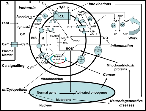
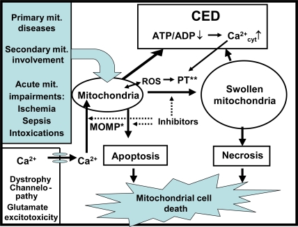
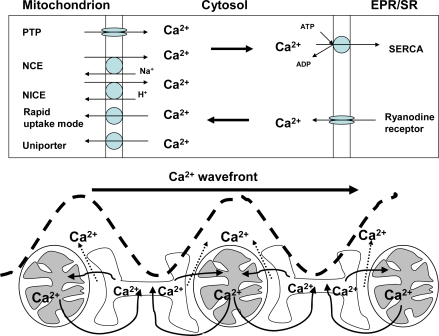
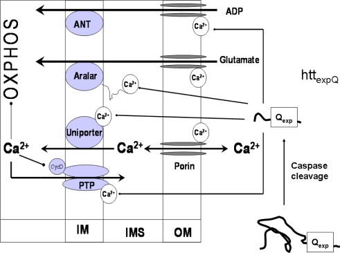
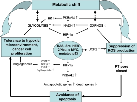
Mitochondrial dysfunction is a hallmark of almost all diseases. Acquired or inherited mutations of the mitochondrial genome DNA may give rise to mitochondrial diseases. Another class of disorders, in which mitochondrial impairments are initiated by extramitochondrial factors, includes neurodegenerative diseases and syndromes resulting from typical pathological processes, such as hypoxia/ischemia, inflammation, intoxications, and carcinogenesis. Both classes of diseases lead to cellular energetic depression (CED), which is characterized by decreased cytosolic phosphorylation potential that suppresses the cell's ability to do work and control the intracellular Ca(2+) homeostasis and its redox state. If progressing, CED leads to cell death, whose type is linked to the functional status of the mitochondria. In the case of limited deterioration, when some amounts of ATP can still be generated due to oxidative phosphorylation (OXPHOS), mitochondria launch the apoptotic cell death program by release of cytochrome c. Following pronounced CED, cytoplasmic ATP levels fall below the thresholds required for processing the ATP-dependent apoptotic cascade and the cell dies from necrosis. Both types of death can be grouped together as a mitochondrial cell death (MCD). However, there exist multiple adaptive reactions aimed at protecting cells against CED. In this context, a metabolic shift characterized by suppression of OXPHOS combined with activation of aerobic glycolysis as the main pathway for ATP synthesis (Warburg effect) is of central importance. Whereas this type of adaptation is sufficiently effective to avoid CED and to control the cellular redox state, thereby ensuring the cell survival, it also favors the avoidance of apoptotic cell death. This scenario may underlie uncontrolled cellular proliferation and growth, eventually resulting in carcinogenesis.

Keywords: cancer; energy depression; hypoxia; inflammation; mitochondria; mitochondrial cell death; neurodegenerative diseases.


Figures

Figure 1.
Figure 1.
The central role of mitochondria in mitochondrial diseases, neurodegenerative diseases, inflammation, ischemia, intoxication and cancer.
Figure 2.
Figure 2.
Mitochondrial cell death. Loss of mitochondrial capacity to synthesize ATP in the processes of OXPHOS leads to cellular energetic depression (CED) characterized by decreased cytosolic phosphorylation potential and increased cytosolic Ca2+ concentration (Ca2+cyt) that leads to reduced ability of cell to do work. The resulting ROS formation and Ca2+ overload further impair the structure and function of mitochondria. In mild stage of CED, when mitochondria can generate some amounts of ATP, mitochondria launch a program of apoptotic cell death by release of cytochrome c. At pronounced CED, when the cytoplasmic ATP levels fall below the levels required for processing the ATP-dependent apoptotic reactions, the cell dies from necrosis. Both, the apoptotic and necrotic death pathways that are mediated by mitochondrial impairments can be classified as of mitochondrial cell death (MCD). The molecular mechanism of mitochondrial outer membrane permeabilization (MOMP) and PT are potential targets for therapeutic interventions preventing mitochondrial cell death.
Figure 3.
Figure 3.
Involvement of mitochondria and endoplasmic/sarcoplasmic reticulum (EPR/SR) in Ca2+ signaling. Upper panel: Mitochondria accumulate Ca2+ via the uniporter and by the rapid uptake mode. Accumulated Ca2+ can be released from mitochondria through reversible Na+ independent (NICE) or Na+ dependent pathways (NCE), but the rates of Ca2+ efflux via these pathways are low in comparison to the fast Ca2+ efflux via the PT pore that can be opened reversibly or irreversibly [46]. Ca2+ accumulation by EPR/SR is realized by SERCA which requires ATP at sufficiently high phosphorylation potentials. To avoid inhibition of SERCA by increasing ADP, it is rephosphorylated by the PCr shuttle (Figure 1). Lower panel: Mitochondria and EPR/SR interact in order to control cytosolic Ca2+ waves and their directed propagation, as modeled in the reconstituted gel system (Table 3 [86]).
Figure 4.
Figure 4.
Mechanisms of regulation of OXPHOS by [Ca2+]cyt that stimulates mitochondrial respiration and ATP synthesis by binding to regulatory sites of several proteins in the mitochondrial outer compartment [40], such as the porin pore [96,98], the PT pore [99], the Ca2+ uniporter, and aralar [94,95]. The Ca2+ binding sites of transporters, PT pore and VDAC may also represent the targets for various pathogenic proteins. As discussed in chapter 3.1.1, huntingtin with an expanded poly Q tract (httexpQ) cleaved by caspases [100,101] can interact with the regulatory Ca2+ binding sites of PT pore and transporters, thereby disturbing the regulation of OXPHOS by [Ca2+]cyt that causes energetic depression, mitochondrial cell death, and tissue atrophy [40].
Figure 5.
Figure 5.
Ca2+-induced inhibition of pyruvate-dependent respiration in isolated muscle mitochondria of transgenic R6/2 HD mice. Multi-substrate inhibitor titration of respiration of isolated mitochondria from skeletal muscle of wild-type (WT) (A,C,E) and transgenic mice (htt150Q) (B,D,F) at the age of 14 to 16 weeks. Isolated muscle mitochondria (0.5 mg/mL) were incubated with 10 mM pyruvate and 2 mM malate. Additions: 10 or 20 μM Ca2+ as indicated; ADP, 2 mM ADP; R, 20 μM rotenone; S, 10 mM succinate; CAT, 10 μM CAT. Thin lines indicate the oxygen concentration in the oxygraph (left ordinate) whereas thick lines represent the rate of respiration in nmol O2/min/mg mitochondrial protein (right ordinate). The height of peaks correlates with the rate of respiration. State 3pyr respiration was adjusted by addition of ADP. Rotenone, an inhibitor of complex I, completely inhibited this respiration. Subsequently, succinate addition allowed the measurement of state 3suc respiration. Due to addition of carboxyatractyloside (CAT), the adenine nucleotide translocator (ANT) was inhibited and the state 4 could be measured. Note, that the pyruvate peak (state 3pyr) is absent in the presence of 20 μM Ca2+ in HD mitochondria. Further details see [139].
Figure 6.
Figure 6.
Mechanisms of mitochondrial involvement in cancer development. Activation of oncogenes and HIF-1α, a typical feature of cancer cell, is associated with downregulation of OXPHOS, its coupling to glycolysis via HKII and upregulation of UCP2 which suppress generation of ROS in mitochondria. This change together with altered balance between anti- and pro-apoptotic genes at mitochondrial membranes decreases the susceptibility of cells to apoptotic death. On the other hand, proliferation and survival of cancer cells is promoted by glycolysis and angiogenesis, both activated by HIF-1α.

Similar articles

Cited by

References

    1. Warburg O, Geissler AW, Lorenz S. Genesis of tumor metabolism by vitamin B1 deficiency (thiamine deficiency) Z. Naturforsch. B. 1970;25:332–333. - PubMed
    1. Luft R. Luft's disease revisited. Severe hypermetabolism of nonthyroid origin with a defect in the maintenance of mitochondrial respiratory control. Mt. Sinai J. Med. 1992;59:140–145. - PubMed
    1. Holt IJ, Harding AE, Morgan-Hughes JA. Deletions of muscle mitochondrial DNA in patients with mitochondrial myopathies. Nature. 1988;331:717–719. - PubMed
    1. Wallace DC, Singh G, Lott MT, Hodge JA, Schurr TG, Lezza AM, Elsas LJ, Nikoskelainen EK. Mitochondrial DNA mutation associated with Leber's hereditary optic neuropathy. Science. 1988;242:1427–1430. - PubMed
    1. Triepels RH, Van Den Heuvel LP, Trijbels JM, Smeitink JA. Respiratory chain complex I deficiency. Am. J. Med. Gen. 2001;106:37–45. - PubMed

Publication types

MeSH terms

Substances