Review
. 2009:542:565-605.
doi: 10.1007/978-1-59745-561-9_30.

Newcastle disease virus: a promising vector for viral therapy, immune therapy, and gene therapy of cancer

Affiliations
Review

Newcastle disease virus: a promising vector for viral therapy, immune therapy, and gene therapy of cancer

Volker Schirrmacher et al. Methods Mol Biol. 2009.

Abstract

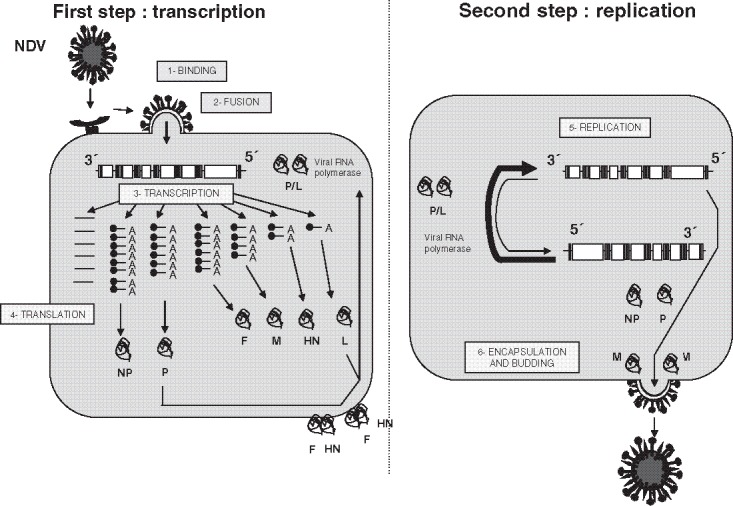
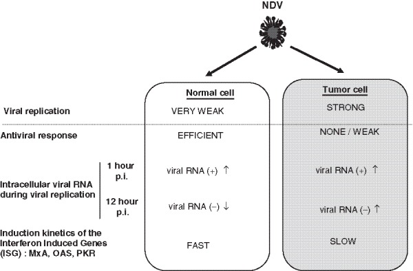
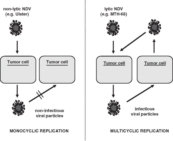
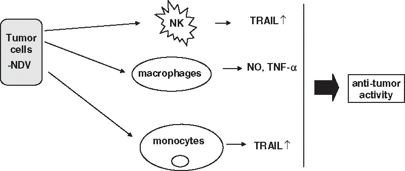
This review deals with the avian paramyxovirus Newcastle disease virus (NDV) and describes properties that explain its oncolytic activity, its tumor-selective replication behavior, and its immune-stimulatory capacity with human cells. The strong interferon response of normal cells upon contact with NDV appears to be the basis for the good tolerability of the virus in cancer patients and for its immune stimulatory properties, whereas the weak interferon response of tumor cells explains the tumor selectivity of replication and oncolysis. Various concepts for the use of this virus for cancer treatment are pointed out and results from clinical studies are summarized. Reverse genetics technology has made it possible recently to clone the genome and to introduce new foreign genes thus generating new recombinant viruses. These can, in the future, be used to transfer new therapeutic genes into tumors and also to immunize against new emerging pathogens. The modular nature of gene transcription, the undetectable rate of recombination, and the lack of a DNA phase in the replication cycle make NDV a suitable candidate for the rational design of a safe and stable vaccine and gene therapy vector.


Figures

Fig. 1
Fig. 1
Cell infection by NDV. NDV replication is composed of two steps (left). The first step consists, after binding and fusion of the virus with the target cell, in the transcription of the viral genes (coding for the nucleoprotein [NP], the phosphoprotein [P], the matrix protein [M], the hemagglutinin–neuraminidase protein [HN], and the large [L] proteins) and in their transaction (right). The second step corresponds to the amplification of the viral genome. The antigenome RNA is used as template for the synthesis of the new viral genomes. The viral RNA-dependent RNA polymerase is composed of the association of the two viral proteins P and L. Encapsulation of the viral genomes occurs at the plasma membrane, from which new virus particles are released via budding from the infected cells (for more details, see the main text, (33), and (167)).
Fig. 2
Fig. 2
Observed differences between interaction of NDV with normal and tumor cells. NDV shows a different pattern of replication in normal cells (left) when compared with tumor cells (right). Its weak replication in normal cells can be correlated with an efficient antiviral response within the infected cells. In contrast, its efficient replication in tumor cells is linked to a weak antiviral response of the latter. These data are based on the analysis of the evolution of the level of genomic (negative) and antigenomic (positive) RNA within the cells during infection (47). One hour after infection, no difference can be observed between normal and tumor cells (47). Twelve hours after infection, the amount of genomic viral RNA was lower in normal cells than in tumor cells (47). The interferon-induced genes (ISG) were also induced differently in normal and tumor cells (for more details, please refer to (47)).
Fig. 3
Fig. 3
Monocyclic versus multicyclic replication patterns of NDV in tumor cells. Left: Non-lytic NDV (for example, the strain Ulster), when added onto a monolayer of tumor cells, leads to the production of viral particles that cannot infect other tumor cells: abortive monocyclic replication. Right: In contrast, lytic NDV strains (for example, the strain MTH-68) lead, after infection of tumor cells, to the production of infectious particles that can infect other tumor cells, thereby leading to an amplification of the viral load: a multicyclic replication cycle.
Fig. 4
Fig. 4
Interactions of NDV with cells of the innate human immune system. NDV, when present at the surface of infected tumor cells, interacts with cells of the innate immunity. It induces the expression of TRAIL at the surface of NK cells and monocytes. It leads also to the production of nitric oxide (NO) and tumor necrosis factor-α (TNF-α) by macrophages. This enhances antitumor immunity (for more details, see the main text).
Fig. 5
Fig. 5
Interactions of NDV with cells of the adaptive human immune system. NDV induces the activation of dendritic cells (DC) that may result in the enhancement of uptake and presentation of tumor-associated peptides loaded on major histocompatibility complex (MHC) molecules to T cells (63). The viral HN protein, via its interaction with T cells, induces costimulatory stimuli (69), thereby activating already existing memory T cells specific for the tumor cells (63). We observed also an increase of TRAIL expression on T cells (100) and a Th1 polarization of the antitumoral response (for more details, see the main text and the references , , and 61).
Fig. 6
Fig. 6
Primordial role of interferon-α induced by NDV at the interface of the innate and adaptive immunity. The interaction of NDV infected tumor cells with the innate immunity leads to the production of high level of interferon-α (especially via the plasmacytoid dendritic cells (PDC)). IFN-α has a central role at the interface between the innate and the adaptive immunity.
Fig. 7
Fig. 7
Rationale for combining TAAs with danger signals in a tumor vaccine. An autologous tumor vaccine modified by infection with NDV (ATV-NDV) consists of tumor cells that are obtained by short in vitro culture of tumor cells from resected tumors of patients. For vaccine production, the cells are infected with the non-lytic NDV strain Ulster and inactivated by γ-irradiation. The use of autologous tumor cells allows the inclusion of individually unique TAAs and also the restimulation of a broad polyclonal antitumor memory response. The modification of the tumor cells by infection with NDV introduced danger signals, co-stimulatory signals, and immunostimulatory properties. For vaccination, 10 million ATV-NDV cells are injected intradermally.
Fig. 8
Fig. 8
Modification of the properties of tumor cells by NDV infection. The replication of NDV in tumor cells leads to the modification of the tumor cells properties and consequently to their interactions with innate (left) and adaptive (right) immunity. Induction of chemokines (such as RANTES, IP-10) and induction of apoptosis by the viral replication stimulate antitumor innate immunity. Viral infection of the tumor cells also leads to enhancement of antigen presentation (notably by upregulation of the expression of MHC molecules) and to enhanced adhesion and co-stimulation (as observed by the increased expression of intercellular adhesion molecule (ICAM)-1 and LFA-3 molecules. Interferon induction by the infected tumor cells also plays an important role in the induction of an antitumor immune response.
Fig. 9
Fig. 9
Tumor neutralization assay: principle (A) and results (B and C). A: Effector cells (PBMC) from a healthy donor are incubated for 5–6 days on top of a tumor cell monolayer with virus-infected tumor vaccine from an established tumor cell line. Activation of effector cells leads to tumor cell killing and to the inhibition of tumor cell growth. This can be quantified by staining the remaining live cells with the chemical reagent MTS. Graphs on the right side show representative data of tumor growth inhibition of mammary carcinoma (B) and stomach carcinoma (C) cells achieved with PBMC from four different healthy donors (for more details, see (147)).
Fig. 10
Fig. 10
Second-generation ATV-NDV tumor vaccine loaded with new immunostimulatory fusion proteins anchored to HN. The ATV-NDV vaccine interacts with T cells via the expression of the tumor antigen (TA) in the context of MHC restriction (signal 1) but also via the expression of the viral HN protein (signal 2). The released IFN-α provides a survival signal to memory T cells represented as signal 3. The addition of the bispecific protein bsHN-CD3 enhances signal 1. The bispecific protein bsHN-CD28 leads to an increased signal 2 (co-stimulation). This optimized tumor vaccine has been showed in in vitro TNA assays to augment T cell antitumor activity as shown in Fig. 11TAA, tumor associated antigen; MHC, major histocompatibility complex; TCR, T cell receptor; IFN-R, interferon type I receptor; PDC, plasmacytoid dendritic cells.
Fig. 11
Fig. 11
Results of tumor neutralization assays showing the superiority of the vaccine ATV-NDV loaded with the two bispecific antibodies bsHN-CD3 and bsHN-CD28. The TNA assays were performed as described in Fig. 9a. Tumor growth inhibition of a mammary carcinoma (A) and a stomach carcinoma (B) cell line with effector cells (from four different healthy donors) activated with tumor vaccine generated from the corresponding mammary carcinoma or the stomach carcinoma cell lines. Notice the increase of antitumor activity upon addition of the bispecific antibody fusion proteins bsHNxCD3, recombinant bispecific single-chain antibodies with a binding specificity for the CD3 on human T cells and also for the viral target molecule HN, which is expressed at the surface of ATV-NDV; bsHNxCD28, recombinant bispecific single-chain antibodies with a binding specificity for the CD28 antigen on human T cells and also for the viral target molecule HN, which is expressed at the surface of ATV-NDV.
Fig. 12
Fig. 12
Incorporation of GM-CSF as therapeutic gene into recombinant NDV. Using reverse genetics, the GM-CSF gene has been inserted before the NP gene within the viral genome. Each viral gene is separated by a junction that consist of three elements, known as gene-end (GE), intergenic (IG), and gene start (GS) sequences. These sequences have been taken into consideration for the insertion of the therapeutic gene. The use of such recombinant virus (rec[GM-CSF]) has been shown to induce an increased antitumor activity when compared with the recombinant virus having no inserted therapeutic gene. The production of GM-CSF after infection of the irradiated tumor cell vaccine was shown to synergize with the virus effect and to lead to an increased production of interferon-α in vitro upon co-incubation with human PBMC (for more details, see (31)). DC, dendritic cells; GM-CSF, growth macrophage colony-stimulating factor; Rec(GM-CSF), recombinant NDV with GM-CSF as insert

Similar articles

Cited by

References

    1. Aghi M., Martzua R.L. Oncolytic viral therapies—the clinical experience. Oncogene. 2005;24:7802–7815. doi: 10.1038/sj.onc.1209037. - DOI - PubMed
    1. Chlichlia K., Schirrmacher V., Sandaltzopoulos R. Cancer immunotherapy: battling tumors with gene vaccines. Curr. Med. Chem. Anti-inflammatory Anti-allergy Agents. 2005;4:353–365. doi: 10.2174/1568014054546290. - DOI
    1. Schirrmacher V. T cell mediated immunotherapy of Metastases: State of the art in (2005) Expert Opin. Biol. Ther. 2005;4(8):1051–1068. doi: 10.1517/14712598.5.8.1051. - DOI - PubMed
    1. Sinkovics J., Horvatz J. New developments in the virus therapy of cancer: a historical review. Intervirology. 1993;36:193–214. - PubMed
    1. Asada T. Treatment of human cancer with mumps virus. Cancer. 1974;34(6):1907–1928. doi: 10.1002/1097-0142(197412)34:6<1907::AID-CNCR2820340609>3.0.CO;2-4. - DOI - PubMed

Substances